M agnetic Resonance Imaging and DW I Features of Orbital Rhabdom yosarcoma
2014-09-17XuetaoMuHongWangYueyueLiYuwenHaoChunnanWuLinMa
Xuetao Mu, Hong Wang, Yueyue Li, Yuwen Hao, Chunnan Wu, Lin Ma
1 Department of Radiology, Chinese PLA General Hospital, Beijing 100853, China
2 Department of MRI, General Hospital of Armed Police, Beijing 100039, China
3 Institute of Orbital Disease, General Hospital of Armed Police, Beijing 100039, China
4 Computer Centre, General Hospital of Armed Police, Beijing 100039, China
Introduction
Materials and methods
Results
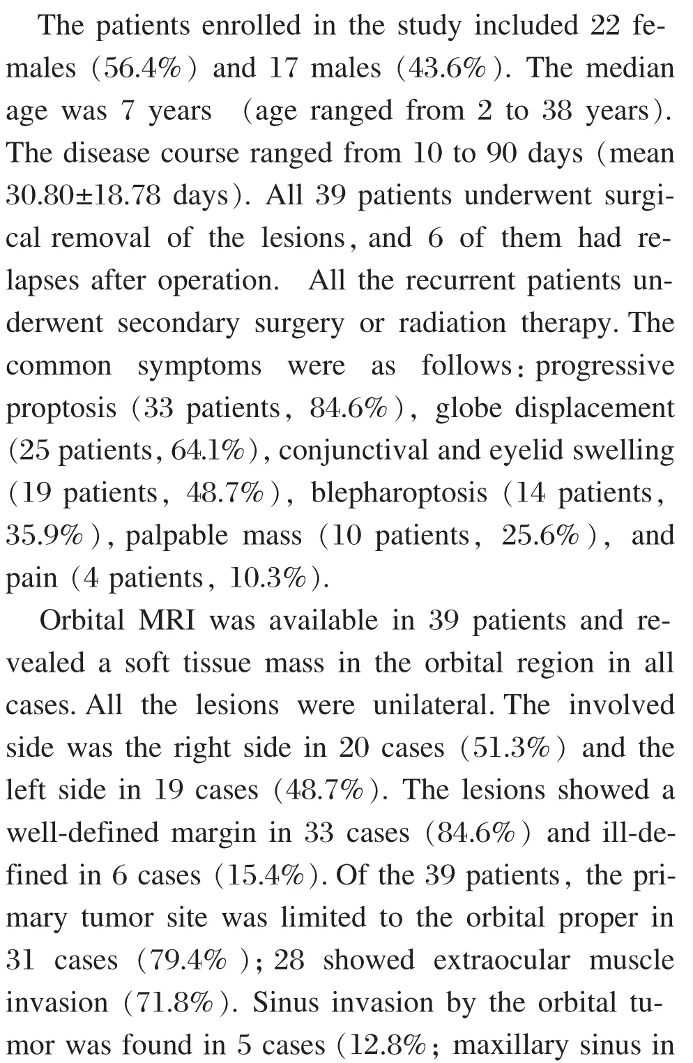
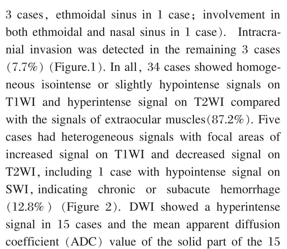
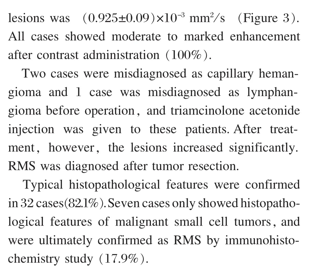
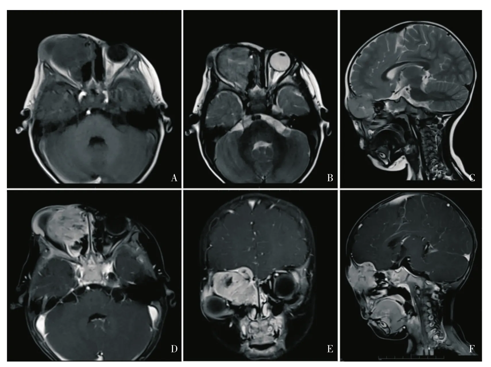
Figure 1 A 2-year-old boy w ith embryonal rhabdomyosarcoma w ith ethmoidal sinus and intracranial invasion.Figure 1A-C:Axial T1W I,T2W I and sagittal T2W I demonstrated a superonasal lesion in the right orbit w ith ethmoidal sinus and intracranial invasion.Figure 1D-F: The lesion and the invaded meninges showed marked heterogeneous enhancement on postcontrast axial, coronal,and sagittal T1WIw ith fat saturation.
Discussion

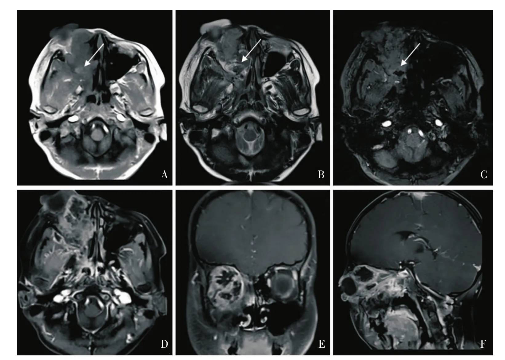
Figure 2 A 10-year-old boy with heterogeneous rhabdomyosarcoma w ith nasal invasion.Figure 2A-B:Axial T1WI and T2WI demonstrated a lesion w ith heterogeneous signals w ith focal areas of increased signal on T1W I and decreased signal on T2W I(arrow).The lesion also showed nasal invasion.Figure 2C: The lesion showed focal areas w ith hypointense signal on SW I(arrow).Figure 2D-F: The lesion showed heterogeneous enhancement on postcontrast axial, coronal, and sagittal T1W I w ith fat saturation.
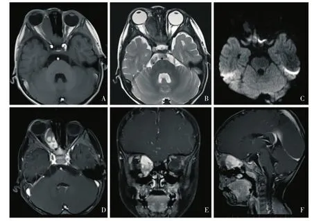
Figure 3 A 2-year-old boy w ith embryonal rhabdomyosarcoma..Figure 3A:.Axial T1W I demonstrated an oval extraconal lesion in the right orbit w ith an isointense signal relative to the muscle..Figure 3B:.The lesion showed a heterogeneously hyperintense signal on T2W I..Figure 1C:.The lesion showed a hyperintense signal on DW I (b=1000)..Figure 3D-F:.The lesion showed a marked heterogeneous enhancement on postcontrast axial, coronal, and sagittal T1W I w ith fat saturation.

杂志排行
眼科学报的其它文章
- A Rat M odel of Autologous Oral M ucosal Epithelial Transplantation for Corneal Limbal Stem Cell Failure
- Clinical Analysis of the Incidence and the Treatment of Pediatric Cataract Patients w ith Optic-nerve M aldevelopment
- Choroidal Analysis of Polypoidal Choroidal Vasculopathy by Spectral Domain Optical Coherence Tomography
- Application of High-frequency Electrosurgical Scalpel and M ethylene Blue Staining in EndonasalDacryocystorhinostom y
- Iontophoretic Delivery of Riboflavin into the Rabbit Cornea:a Primary Study
- Tendency for Evolution of High M yopia in 308 Chinese School Children from Xi'an City
