Effects of HLEC on the secreted proteins of epithelial ovarian cancer cells prone to metastasize to lymph nodes
2013-09-26XinYingZhangFuQiangYinLiLiuTingGaoHeYunRuanXiaoGuanYingXinLuDanRongLi
Xin-Ying Zhang, Fu-Qiang Yin, Li Liu, Ting Gao, He-Yun Ruan, Xiao Guan, Ying-Xin Lu, Dan-Rong Li
1The Cancer Hospital Affiliated to Guangxi Medical University, Nanning 530021, China; 2The Experiment Center of The Guangxi Medical University, Nanning 530021, China
Introduction
The delitescent onset of epithelial ovarian cancer (EOC) is possible.Early metastasis occurs easily, in which lymphatic metastasis is the primary route of metastasis.The mortality rate of EOC is the highest among malignant gynecological tumors.Studies have recently indicated that lymphangiogenesis in tumor is closely related to tumor node metastases, which can be promoted by lymphatic proliferation1.
In the previous study, sublines of human ovarian papillary serous carcinoma cells (SKOV3) in lymphatic metastasis were cloned in nude mice, wherein the fourth generation of SKOV3 (SKOV3-PM4) cells were screened and established with directional highly lymphatic metastasis potential2.In the present study, we conducted experiments on solitary culture and co-cultures of SKOV3, SKOV3-PM4, and human lymphatic capillary endothelial cells (HLEC) to investigate the effect of HLEC on proteins secreted by EOC cells with directional highly lymphatic metastasis (SKOV3-PM4).This study provides a rationale for the therapy of antilymphangiogenesis and anti-lymphatic metastasis among patients with EOC.
Materials and methods
Study object
SKOV3 cell lines were purchased from the Center of Cell Resources of Shanghai Institutes for Biological Sciences, whereas SKOV3-PM4 cell lines were prepared by this research group2.HLEC was purchased from the US Sciencell Co.Sources of serum specimens were obtained from 20 benign ovarian tumor patients and 20 ovarian carcinoma patients with lymph node metastasis.All of the participants were patients with definite pathological diagnosis made by the Cancer Hospital Affiliated to Guangxi Medical University.The patients with ovarian cancer were 32 to 60 years of age, and the median age was 52.They were not surgically and chemo radio therapeutically treated before blood specimen collection.A total of 20 healthy women (normal physical examinees; 38 to 55 years old; median age was 43) were included in the control group.The participants in the control group had no history of infection and medication within four weeks before the study.The institute received approval of the Ethics Committee as well as consent from the patients.
Primary reagents
The cytokine antibody array was purchased from the US RayBiotech Co, and the ITRAQ kit was from the US ABI company.The ELISA kit was purchased from the American Abcam Co.Special culture medium (ECM) for HLEC were purchased from the US Sciencell Co., HPLC (1,200 series) from Agilent Co.,and Nano-LC separation point target system and 5800 MALDITOF/TOF protein analyzer were from the American ABI Co.
Preparation of supernatant from cell culture
Three groups of cells, namely, SKOV3, SKOV-PM4, and HLEC,were digested at the logarithmic growth phase (106cells).For co-cultures, 5×10⁵HLEC cells were added to culture flasks containing the same amount of SKOV3 and SKOV3-PM4 cells.After 24 h, the medium was replaced with basal ECM without serum and cytokine.After 48 h, the supernatant from the cell cultures were collected.The supernatants were then refrigerated at –80 °C after dividing them into different groups, namely,SKOV3 (A), SKOV3+HLEC (B), SKOV3-PM4 (C), SKOV3-PM4+HLEC (D).
Detection of cytokine antibody array
The procedure was conducted according to the RayBio human cytokine 507 antibody array manual.The cell supernatants were labeled with biotin.The array was balanced and then closed.The specimens were cleansed, incubated with fluorescerstreptavidin, and cleansed again.Images were then obtained using a Uniscand1000 scanner.The data were processed with a Scanalyze software.Standard values of individual antibody expression were then calculated.Cytokines with differential expressions were screened according to the following criteria:
Upregulation: the ratio of group C/A and D/C ≥1.33.Downregulation: the ratio of group C/A and D/C ≤0.77.
Detection with ITRAQ-2D-LC-MALDI-TOF/TOF/MS
Proteolysis was performed using ITRAQ labels (113, 114, 115,and 116, which correspond to the four groups of samples marked A, B, C, and D, respectively).Chromatographic separation and targeting were conducted according to reference3and the operation manual of the device.
Mass spectrometry (MS) and database retrieval: A 5800 Plus MALDI-TOF/TOF mass spectrum analyzer was used in tandem with MS (MS/MS) to perform identification and relative quantitative analysis of polypeptides.ProteinPilot 2.0 was used to perform retrieval and identification of proteins from the MS data against the NCBInr database.Relative quantitative analysis was conducted for proteins at a confidence level of ≥95% by using the peak area integrals of ions reported from m/z 114, 115,116, and 117.Results with P≤0.05 were chosen for the report based on the ratio of 115:113 and 116:115.Manual con firmation was not performed for protein polypeptide matches of more than one high confidence level (no less than 99%), whereas manual con firmation was performed to the fragment ions of the MS/MS spectrum of proteins with matching polypeptide without a high con fidence level (no less than 99%).
Bioinformatics analysis
Bioinformatics analysis was conducted on proteins with intersection of differences in array detection and MS.Proteins with significant differences were chosen for verification in human serum specimens and supernatants from cell cultures via the ELISA method.The instruction manual provided by ABCAM Co.for the ELISA kit was strictly followed.
Statistical analysis
All data quantification and satistical analyses were performed using SPSS16.0 software.Data were presented as the mean ± SD.Analysis of variance was performed to compare measurement data.P value <0.05 were considered statistically significant.
Results
Cytokine antibody array detection
By comparing SKOV3-PM4 with SKOV3, 39 differential proteins were found in the SKOV3-PM4 culture supernatant, in which 34 were upregulated and 5 were downregulated.By comparing SKOV3-PM4+HLEC with SKOV3-PM4, 41 differential proteins were found after co-culture, in which 22 were upregulated and 19 were downregulated.The results are shown in Table 1.
ITRAQ label combined with MALDI-TOF-TOF MS/MS
After data analysis, SKOV3-PM4 was compared with SKOV3,and 36 differential proteins were present in the SKOV3-PM4 culture supernatant, in which 22 were upregulated and 14 were downregulated.
By comparing SKOV3-PM4+HLEC with SKOV3-PM4, 65 differential proteins were found in the co-culture, in which 37 were upregulated and 28 were downregulated.The expressions of VEGFA and GRN was upregulated, whereas that of SPARC and IGFBP7 was downregulated.This result was consistent with the tendency of the array, as shown in Table 2.
Bioinformatics analysis conducted on differential cytokines
Bioinformatics analysis was conducted on the screened differential cytokines.The figure of network regulation relationship between the differential proteins and the lymphatic metastasis of EOC was obtained using a Coremine software (Figure 1).
在党中央、国务院的坚强领导下,玉树抗震救灾工作快速有力有序有效地展开,取得了令人瞩目的成绩,赢得了全世界的赞誉。
Figure 1 shows different link thicknesses that represent different correlation degrees among proteins, biological processes, and EOC, as well as lymphatic metastasis.Thicker lines represent higher correlations between both ends of the line.The analysis showed that OEC is closely associated with VEGFA,SPARC, and GRN, but it is not correlated with IGFBP7.Interaction network among IGFBP7 and the three proteins that are closely correlated with ovarian cancer were drawn using GeneMania to analyze the relationship between IGFBP7 and lymphatic metastasis of ovarian cancer.In Figure 2, the lines in different colors represent different correlations, in which the thickness corresponds to the degree of correlation.
Figure 2 shows that coexpression and co-localization are apparent among multiple proteins, such as IGFBP7, SPARC,VEGFA and CXCL10.These proteins have common structural domain and interact with one another.In determining the functions of IGFBP7 and other proteins in terms of their biological behaviors, we can speculate that they are possibly important in metastasis.The functions of the relevant proteins were analyzed using a DAVID software.The results show that the proteins are primarily involved in the regulation of cell migration,cell adhesion, and neovascularization (Table 3).
ELISA verification
Bioinformatic analysis results show that IGFBP7 is closelycorrelated with VEGFA protein, but no scientific reports on IGFBP7 and ovarian cancer are currently available.Therefore,IGFBP7 and VEGFA were selected for ELISA verification in this study.With regard to VEGFA, any pairwise comparison was statistically significant (P<0.05) among the three groups, in which the expression level in the malignant group was the highest.As for IGFBP7, the difference of any pairwise comparison was also statistically significant, in which the tendency was opposite to that of VEGFA, that is, the expression level in the normal group was the highest.In pairwise comparison of the cell supernatants between SKOV3 and SKOV3-PM4 as well as SKOV3-PM4 and SKOV3-PM4+HLEC, the expression tendencies of both proteins were statistically significant (P<0.05), which was similar to array detection and MS (Figures 3,4).

Table 1 Differentially expressed proteins in different cell supernatants

Table 2 Intersection of differential protein expression in the antibody arrays and iTRAQ-2D-LC-MALDI-TOF/TOF/MS

Figure 1 Regulation network of differential proteins and lymphatic metastasis in ovarian cancer.

Figure 2 Function prediction of IGFBP7 by GeneMANIA.
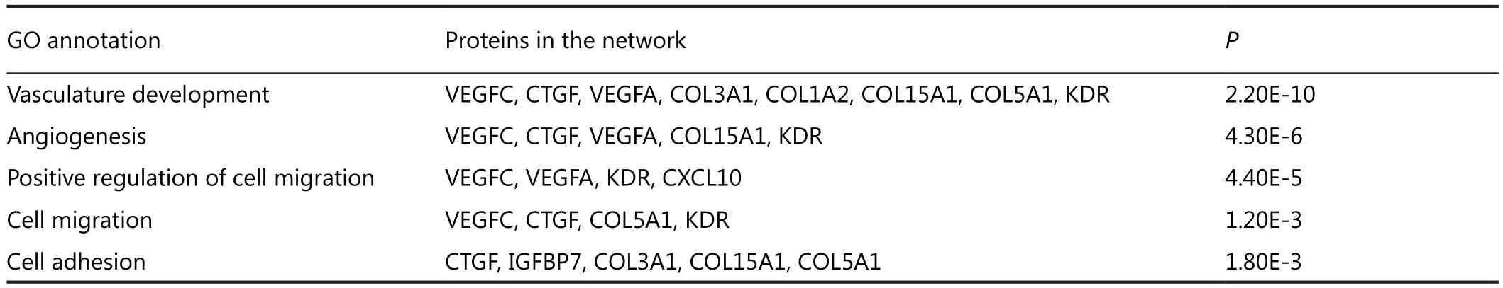
Table 3 The annotated functions of IGFBP7 and other proteins related to metastasis in GeneMANIA network (as known in Figure 2)
Discussion
Numerous studies have shown that after tumor cells contact with HLEC in a tumor microenvironment, metastasis and invasion of tumor cells occurred by inducing lymphangiogenesis to drain the lymph fluid and the tumor cells.The new lymphatic vessels provide nutrients and oxygen to promote or maintain the growth of tumor cells.Thus, lymphangiogenesis is an important pathway for invasion and metastasis of tumors4.Directional lymphatic metastasis of tumor primarily depends on the migration, adherence, and penetration of tumor cells to HLEC.In the present study, HLEC was used to co-culture with SKOV3-PM4 cells.We simulated the tumor environment of HLEC and used the cytokine antibody array for detection.Bioinformatics analysis showed that the upregulation of VEGFA and GRN, as well as the downregulation of IGFBP7 and SPARC, were correlated with lymphatic metastasis of EOC, and that VEGFA was an important node protein.

Figure 3 VEGFA concentrations in serum and cell supernatant.
It is clear that the angiogenesis factor VEGFA is involved in tumor growth and spread, and promotes angiogenesis by binding VEGFR1 and VEGFR2.VEGFR2 is reported to be expressed in HLEC, and VEGFC and VEGFD can bind VEGFR2 and VEGFR3, mediating lymphangiogenesis5.In cursiefen-induced angiogenesis and lymphangiogenesis6in normal cornea without blood and lymph vessels, the function of VEGFA is blocked but that of VEGFC and VEGFD was retained.Angiogenesis and lymphangiogenesis are simultaneously blocked, suggesting that VEGFA simultaneously promotes angiogenesis and lymphangiogenesis.Recently, VEGF-A has been found to function in HLEC by promoting lymphangiogenesis and the lymphatic metastasis of cancer cells.Spannuth7found that the expression level of VEGFR2 in patients with ovarian cancer was high.Qiu8detected the expression of VEGFAmRNA in ovarian cancer by using RT-PCR and immunohistochemistry.The density of the lymph vessels was manually determined, and the results implied that the high expression level of VEGFA is correlated with lymphangiogenesis and lymph node metastasis.
GRN is an autocrine growth factor closely related with the onset and development of various tumors, such as ovarian and esophageal cancer.Liu9tested the expression level of GRN in three types of human ovarian cancer cell lines with different malignant potentialities and demonstrated that the expression level of GRN was closely and positively correlated with tumor cell proliferation and invasiveness.In the classical signal transduction pathway of VEGFA in promoting angiogenesis, GRN is involved in three kinase pathways that regulate tumor growth (MAPK, P13K, and FAK).We speculated that GRN, as an upstream gene protein,might indirectly promote angiogenesis and lymphangiogenesis of tumor by regulating the expression pathway of VEGFA10.

Figure 4 IGFBP7 concentrations in serum and cell supernatant.
SPARC is an extracellular matrix glycoprotein with complex functions and tissue specificity.SPARC in tumor tissues can alter the function of stroma and tumor cells and activate downstream pathways, such as ILK, FAK, and MAPK, especially the expression of relevant factors of angiogenesis, thereby controlling tumor growth and invasion11.In an animal model of ovarian cancer12, the lack of SPARC upregulated the expression of VEGF, thereby promoting angiogenesis, lymphangiogenesis,and the metastatic ability of cancer cells.
In summary, this study showed that more than 41 proteins with different expression are present after co-culture of SKOV3-PM4 with HLEC.According to literature, these proteins are involved in functions related to stroma cells, cell adhesion factors,interleukin, chemotactic factor, and cell signal transduction.Further studies on the specific functions and impacts of these proteins on the onset and development of lymph node metastasis of ovarian cancer are warranted.
Acknowledgements
This work was supported by the National Natural Science Foundation of China (Grant No.81060218) and Guangxi Natural Science Foundation (Grant No.2012GXNSFAA053157).
Conflict of interest statement
No potential conflicts of interest are disclosed.
1.Wang Y, Oliver G.Current views on the function of the lymphatic vasculature in health and disease.Genes Dev 2010;24:2115-2126.
2.Ruan HY, Li DR, Li L, Guan X, Zhang W.Establishment of human ovarian carcinoma cell lines with directional highly lymphatic metastasis and study of their biological characteristics.Zhonghua Fu Chan Ke Za Zhi 2007;42:482-486.
3.Hou Q, Tan HT, Lim KH, Lim TK, Khoo A, Tan IB, et al.Identification and functional validation of caldesmon as a potential gastric cancer metastasis-associated protein.J Proteome Res 2013;12:980-990.
4.Duong T, Koopman P, Francois M.Tumor lymphangiogenesis as a potential therapeutic target.J Oncol 2012;2012:204946.
5.Dellinger MT, Brekken RA.Phosphorylation of Akt and ERK1/2 is required for VEGF-A/VEGFR2-induced proliferation and migration of lymphatic endothelium.Plos One 2011;6:e28947.
6.Cursiefen C, Chen L, Borges LP, Jackson D, Cao J, Radziejewski C,et al.VEGF-A stimulates lymphangiogenesis and hemangiogenesis in inflammatory neovascularization via macrophage recruitment.J Clin Invest 2004;113:1040-1050.
7.Spannuth WA, Nick AM, Jennings NB, Armaiz-Pena GN, Mangala LS, Danes CG, et al.Functional significance of VEGFR-2 on ovarian cancer cells.Int J Cancer 2009;124:1045-1053.
8.Qiu M, Liang ZQ, Wang L.Relationship between expression of vascular endothelial growth factor A and lymphangiogenesis and lymph node metastasis.Shi Yong Yi Xue Za Zhi 2005;21:2474-2476.
9.Liu Y, Xi L, Liao G, Wang W, Tian X, Wang B, et al.Inhibition of PC cell-derived growth factor (PCDGF)/granulin-epithelin precursor (GEP) decreased cell proliferation and invasion through downregulation of cyclin D and CDK4 and inactivation of MMP-2.BMC Cancer 2007;7:22.
10.Diaz-Cueto L, Arechavaleta-Velasco F, Diaz-Arizaga A, Dominguez-Lopez P, Robles-Flores M.PKC signaling is involved in the regulation of progranulin (acrogranin/PC-cell-derived growth factor/granulin-epithelin precursor) protein expression in human ovarian cancer cell lines.Int J Gynecol Cancer 2012;22:945-950.
11.Chlenski A, Cohn SL.Modulation of matrix remodeling by SPARC in neoplastic progression.Semin Cell Dev Biol 2010;21:55-65.
12.Said N, Socha MJ, Olearczyk JJ, Elmarakby AA, Imig JD, Motamed K.Normalization of the ovarian cancer microenvironment by SPARC.Mol Cancer Res 2007;5:1015-1030.
13.Oh Y.IGFBPs and neoplastic models.New concepts for roles of IGFBPs in regulation of cancer cell growth.Endocrine 1997;7:111-113.
14.Persano L, Crescenzi M, Indraccolo S.Anti-angiogenic gene therapy of cancer: current status and future prospects.Mol Aspects Med 2007;28:87-114.
15.Evdokimova V, Tognon CE, Benatar T, Yang W, Krutikov K,Pollak M, et al.IGFBP7 binds to the IGF-1 receptor and blocks its activation by insulin-like growth factors.Sci Signal 2012;5:ra92.
猜你喜欢
杂志排行

Cancer Biology & Medicine的其它文章
- Asian trends in primary androgen depletion therapy on prostate cancer
- Therapeutic resistance in cancer: microRNA regulation of EGFR signaling networks
- Why bortezomib cannot go with ‘green’?
- Analysis of 30 patients with persistent or recurrent squamous cell carcinoma of the cervix within one year after concurrent chemoradiotherapy
- Rare myeloid sarcoma/acute myeloid leukemia with adrenal mass after allogeneic mobilization peripheral blood stem cell transplantation
- Instructions for Authors