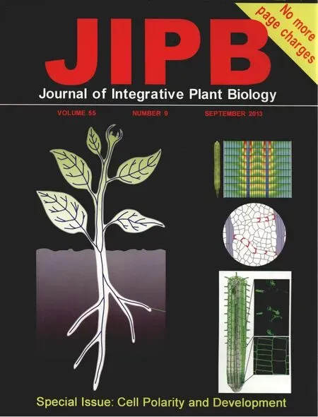
Cell Polarity and Development
2013-02-20RemkoOffringa,JürgenKleine‐Vehn
All cells show some degree of polarity,either by asymmetrically distributed membrane or cytosolic components.Even in bacterial cells that do not have the eukaryotic membrane compartmentalization of the cytoplasm,proteins can be localized at specific areas.In rod‐shaped bacteria,many processes such as signaling,flagella formation,and DNA uptake occur at the cell poles.In addition,the symmetry of cell division in these bacteria is determined by Min proteins,which in Bacillus subtilus are specifically positioned at the cell poles by the DivIVA protein.The cytosolic DivIVA seems to preferentially accumulate at negatively curved membranes,suggesting that membrane curvature is a primary cell polarity determinant(Dacks and Field 2007;Strahl and Hamoen 2012).
Cell polarity has been crucial for the evolution of multicellular organisms,as it is at the basis of asymmetric(formative)cell division,which is an essential step for cellular differentiation,enabling the daughter cells to acquire a different cell fate.A key example of transiition to multicellularity is provided by the green algea Volvox carteri,where asymmetric cell division leads to the formation of small vegetative cells and large generative conidia(Nishii and Miller 2010).Especially in higher plants,formative cell divisions are used during many stages of development.This is important because,unlike animal cells,plant cells are embedded in the extracellular matrix of the cell wall,which fixes their position and prevents their differentiation by migrating to new morphogen fields.Polarization in plant development starts with the first cell division,where the zygote asymmetrically elongates and divides into a smaller apical cell that will develop into the embryo proper,and a larger cell that only divides longitudinally to form a single cell file called the suspensor(Ueda and Laux 2012).Asymmetric cell divisions occur every time a new cell layer or cell type has to be established.A nice example of developmentally important asymmetric cell division is the formation of stomatal lineage cells by asymmetric division of the meristemoid mother cell.Further asymmetric division of stomatal lineage cells guarantees that upon their terminal differentiation the stomata are nicely spaced over the leaf surface with at least one pavement cell in between to guarantee efficient gas exchange for the photosynthesizing cells.Molecular evidence for cellular polarization was provided in this specific case by the identification of the BASL protein,which shows a very dynamic localization,switching from the nucleus to the plasma membrane,where it marks the side of the cell giving rise to the larger daughter cell,eventually differentiating into a pavement cell(Dong et al.2009;Robinson et al.2011).
Another aspect of cell polarity is of course the asymmetric growth of cells,which is exemplified by lobe pattern formation of pavement cells,and is drawn to the extreme in tip growing cells such as root hairs,trichomes and pollen tubes(Xu et al.2010;Rounds and Bezanilla 2013).Thus,cell polarity is crucial from the start to the final stages of plant development.
Cell polarity is also important for polar transport processes that induce asymmetric distribution of signaling molecules directing plant development,such as the plant hormones auxin and gibberellin(Grunewald and Friml 2010;Shani et al.2013).
In general,cell polarity is determined by the asymmetric distribution of cell components.One of the key questions is what determines the compartmentalization and distribution of these cell components?From the bacterial DivIVA example it is clear that asymmetrical distribution of cytosolic proteins can be self‐organizing,driven by the shape of the cell membrane and the 3D structure of the protein,thereby providing attachment points for other proteins(Strahl and Hamoen 2012).In larger eukaryotic cells,the cytoskeleton is believed to play a central role in asymmetric distribution of cell components by mediating the directional trafficking of membrane components and their cargo.However,there are also models suggesting that many of these trafficking processes would occur in the absence of cytoskeletal components.This indicates how little we know about cellular polarization mechanisms.
In this special issue of JIPB several aspects of plant cell polarity are highlighted.Not surprisingly,in view of its central role in plant development,the mechanism of polar transport of the hormone auxin is discussed in several of the contributions.We start with basic processes that determine the asymmetric distribution of plasma membrane(PM)proteins,being their post‐translation modification.In the first two reviews,the authors summarize the basic components that determine cell polarity,and stress the importance of sorting signals in PM proteins and how post‐translational modification by phosphorylation and ubiquitination directs the trafficking of these PM proteins(Korbei and Luschnig 2013;Offringa and Huang 2013).In plants,relatively little is known about how these processes are utilized,and the only example where detailed studies have uncovered the entire complex from kinase to phosphorylation sites and also the ubiquitination sites are the PIN auxin efflux carriers.For phosphorylation,a comparison with some examples from animal cells is made in order to provide clues as to how phosphorylation of PM proteins could act on their sorting.As expected,phosphorylation results in the recruitment of PM proteins into specific protein complexes involved in trafficking,and these can be either adaptor complexes related to clathrin‐mediated vesicle trafficking,or cytoskeleton‐interacting proteins.
The review by Barbara Korbei and Christian Luschnig(2013)covers ubiquitination of PM proteins,and how this affects their trafficking and may lead to their degradation.The ESCRT‐0 to ‐III and retromer complexes are the main components involved in animal cells;however,although similar components have been identified in plants,their role is largely unknown.The review also nicely demonstrates how PM protein ubiquitination alters plant development.
Subsequently,Megan Sawchuck and Enrico Scarpella(2013)discuss how the polarity of auxin transport is required to mediate vascular development.The authors integrate the long‐standing “auxin canalization hypothesis”with recent data addressing the polarized alignment of vascular strands(Sawchuk and Scarpella 2013).Development is also directed by the polar delivery of cell wall components,and this is the subject of the review by Fangwei Gu and Erik Nielsen,who review this topic by looking at tip growth,where this polar delivery process is obviously superimposed(Gu and Nielsen 2013).The authors particularly highlight the crucial mechanisms for defined cell wall deposition and its role in controlled,polar cellular expansion.
All of this basic knowledge of cell polarity regulation is required to understand complex developmental processes,and we start to reveal how these processes eventually shape entire organs.Verônica Grieneisen and coauthors touch upon this subject by presenting the development of one of the most complex organs of a plant,the gynoecium(Grieneisen et al.2013).The review nicely demonstrates how mathematical modeling can be used to simulate cell polarity dynamics during complex developmental processes such as gynoecium development,and how this leads to new insights.Finally,the research paper by Christian Löfke and coauthors presents a detailed study on the polar growth of the root epidermal cell layer,which consists of two files of cells called the tricho‐and the atrichoblasts(Löfke et al.2013).Trichoblasts develop polar outgrowth in the differentiation zone,leading to root hair formation.In contrast,the atrichoblasts give rise to non‐hair cells.Tricho‐and atrichoblast show distinct cell size determination in the root meristem,giving rise to smaller and bigger cell files(Galway et al.1994;Berger et al.1998;Dolan 2001).Since both cell files need to support uniform growth of the root epidermis,the growth of the independent cell files must be coordinated,and the authors provide evidence that this coordination is mediated via an interdependent,non‐cell autonomous mechanism.
We would like to express our gratitude to the authors for their excellent contributions,which we anticipate to provide you as reader of JIPB with a solid basis on plant cell polarity and development.
Remko Offringa
Issue Editor
Molecular and Developmental Genetics
Institute Biology Leiden
Leiden University
Sylvius Laboratory
Sylviusweg 72
2333 BE The Netherlands
Jürgen Kleine‐Vehn
Issue Editor
Universität für Bodenkultur Wien(BOKU)
Institute of Applied Genetics and Cell Biology(IAGZ)
Muthgasse 18
1190 Wien
Austria
Berger F,Hung CY,Dolan L,Schiefelbein J(1998)Control of cell division in the root epidermis of Arabidopsis thaliana.Dev.Biol.194,235–245.
Dacks JB,Field MC(2007)Evolution of the eukaryotic membrane‐trafficking system:Origin,tempo and mode.J.Cell Sci.120,2977–2985.
Dolan L(2001)How and where to build a root hair.Curr.Opin.Plant Biol.4,550–554.
Dong J,MacAlister CA,Bergmann DC(2009)BASL controls asymmetric cell division in Arabidopsis.Cell 137,1320–1330.
Galway ME,Masucci JD,Lloyd AM,Walbot V,Davis RW,Schiefelbein JW(1994)The TTG gene is required to specify epidermal cell fate and cell patterning in the Arabidopsis root.Dev.Biol.166,740–754.
Grieneisen VA,Marée AFM, Østergaard L(2013)Juicy stories on female reproductive tissue development:Coordinating the hormone flows.J.Integr.Plant Biol.55,847–863.
Grunewald W,Friml J(2010)The march of the PINs:Developmental plasticity by dynamic polar targeting in plant cells.EMBO J.29,2700–2714.
Gu F,Nielsen E(2013)Targeting and regulation of cell wall synthesis during tip growth in plants.J.Integr.Plant Biol.55,835–846.
Korbei B,Luschnig C(2013)Plasma membrane protein ubiquitylation and degradation as determinants of positional growth in plants.J.Integr.Plant Biol.55,809–823.
Löfke C,Dünser K,Kleine‐Vehn J(2013)Epidermal patterning genes impose non‐cell autonomous cell size determination and have additional roles in root meristem size control.J.Integr.Plant Biol.55,864–875.
Nishii I,Miller SM(2010)Volvox:Simple steps to developmental complexity?Curr.Opin.Plant Biol.13,646–653.
Offringa R,Huang F(2013)Phosphorylation‐dependent trafficking of plasma membrane proteins in animal and plant cells.J.Integr.Plant Biol.55,789–808.
Robinson S,Barbier deRP,Chan J,Bergmann D,Prusinkiewicz P,Coen E(2011)Generation of spatial patterns through cell polarity switching.Science 333,1436–1440.
Rounds CM,Bezanilla M(2013)Growth mechanisms in tip‐growing plant cells.Annu.Rev.Plant Biol.64,243–265.
Sawchuk MG,Scarpella E(2013)Polarity,continuity and alignment in plant vascular strands.J.Integr.Plant Biol.55,824–834.
Shani E,Weinstain R,Zhang Y,Castillejo C,Kaiserli E,Chory J,Tsien RY,Estelle M(2013)Gibberellins accumulate in the elongating endodermal cells of Arabidopsis root.Proc.Natl.Acad.Sci.USA 110,4834–4839.
Strahl H,Hamoen LW(2012)Finding the corners in a cell.Curr.Opin.Microbiol.15,731–736.
Ueda M,Laux T(2012)The origin of the plant body axis.Curr.Opin.Plant Biol.15,578–584.
Xu T,Wen M,Nagawa S,Fu Y,Chen JG,Wu MJ,Perrot‐Rechenmann C,Friml J,Jones AM,Yang Z(2010)Cell surface‐and rho GTPase‐based auxin signaling controls cellular interdigitation in Arabidopsis.Cell 143,99–110.
