足部常见软组织肿物的MRI诊断
2012-11-07张朝晖方字文
张朝晖,方字文
足部软组织肿物多为良性,恶行者很少见;有研究显示足部软组织肿瘤中良性者占87%[1]。MRI对软组织病变的显示能力强,但有关足部软组织肿物MRI表现的报道尚少见,笔者对足部常见软组织肿物的MRI特征及有关临床和病理特点进行总结分析。
1 海绵状血管瘤
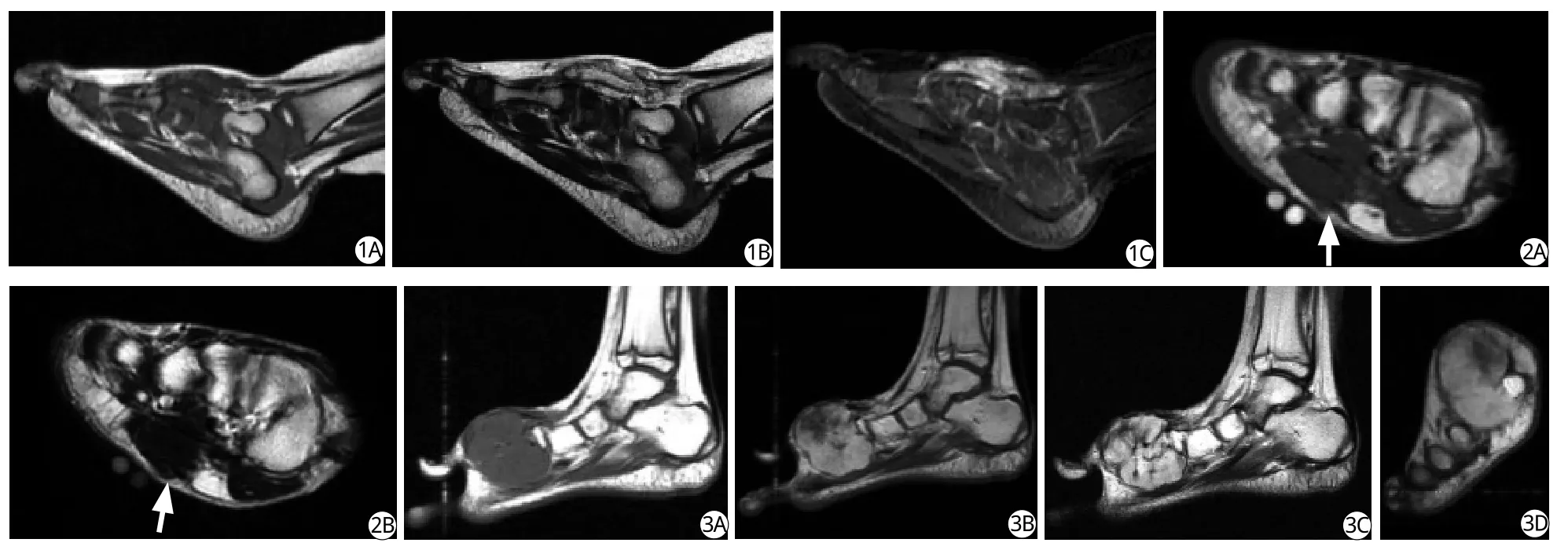
足部海绵状血管瘤较常见,可累及皮肤、皮下和深部组织。组织学上,海绵状血管瘤除了血管成分外,还有脂肪、纤维、平滑肌、含铁血黄素、血栓等非血管成分;其内还常有钙化,50%在平片上可见静脉石[2]。位置表浅者查体可触及质软肿物,并可见局部皮肤发紫。海绵状血管瘤在T2WI多表现为分叶状高信号病灶,且信号强度随T2权重的增加而增高,这主要由病变内的缓慢流动的血液引起;另外脂肪成分在T2WI也呈较高信号;含铁血黄素、流空的血管、钙化及纤维组织在T2WI呈低信号;病变内亚急性出血和脂肪成分在T1WI呈高信号表现;注射对比剂后病变多呈较明显强化[2-3](图1)。
2 跖腱膜纤维瘤病
跖腱膜纤维瘤病是足底腱膜的非肿瘤性纤维结节样增生,早期病变内的梭形纤维母细胞成分较多伴有一定数量的胶原成分;后期胶原成分增多,细胞成分减少[4]。它可与掌腱膜纤维瘤病和阴茎纤维瘤病等其他浅表纤维瘤病同时存在,可呈单发或多发结节,多位于足底腱膜的内侧浅表部分[2,4]。
本病多见于30岁以上患者,男性发病多于女性,临床上可见质硬皮下结节,长期站立或行走后疼痛,是足跟部疼痛最常见的原因之一,在长期跑步者和肥胖者尤其多见[2,5]。病变在T1WI和T2WI的信号强度等于或低于邻近的肌肉[4](图2),增强扫描呈轻度或明显强化[5],并可伴有跟骨骨刺和皮下组织水肿[2]。
3 侵袭性纤维瘤病
侵袭性纤维瘤病起源于肌肉的结缔组织及其表面的筋膜和腱膜,可包裹肌肉和肌腱,又称为肌腱膜纤维瘤,韧带状纤维瘤[4-6]。它由分化较好的纤维母细胞和肌纤维母细胞增生形成,胶原纤维穿插于细胞之中,多少不等[6]。
本病常发生于10~50岁年龄段,尤其多见于25~35岁,常表现为深在、边界不清的肿块,疼痛多不明显[5]。其生物学行为介于纤维瘤和纤维肉瘤之间,一般累及范围较广泛,但少见多中心发病,切除后易复发,但无转移,恶变为肉瘤也罕见[4-5,7]。足部是其好发部位之一[4]。
在MRI上侵袭性纤维瘤病表现为形态不规则的肿块,常累及多块肌肉,并可沿着筋膜呈线带状蔓延,无包膜或包膜不完整,边界可不清,瘤内一般无坏死,瘤周一般无水肿,邻近骨可受压,但髓腔一般不受侵犯;在T1WI以等、低信号为主,在T2WI以高信号为主,瘤内可见带状或条片状与肌肉相比呈等或低信号的区域;注射对比剂后,肿瘤呈中等程度或明显强化,其内与肌肉相比呈等或低信号的区域不强化或轻度强化[5-6](图3)。

图1 足背海绵状血管瘤。A:矢状面T1WI示足背肿物,与肌肉相比呈等信号,混杂斑片状高信号成分。B:矢状面T2WI示肿物以高信号为主,混杂少量条点状低信号。C:矢状脂肪抑制增强T1WI示肿物不均匀明显强化 图2 足底跖腱膜纤维瘤病。冠状T1WI (A)、T2WI (B)示足底腱膜结节样增厚(箭),病灶与肌肉相比病变在T1WI和T2WI呈等信号,邻近皮下脂肪受压 图3 足部侵袭性纤维瘤病。A:矢状面T1WI示足内侧部分叶状肿物,与肌肉相比呈等信号。B:矢状面T2WI示肿物主要呈高信号,混杂条片状低信号成分。C:矢状面增强T1WI示肿物明显强化,其内T2WI等低信号成分无或轻度强化。D:冠状面增强T1WI示第一跖骨被肿物包绕,但未见明显骨质破坏Fig. 1 Cavernous hemangioma in the dorsal aspect of foot. A: Sagittal T1-weighted MR image shows a soft tissue mass in the dorsal aspect of foot, which is predominantly isointense relative to skeletal muscle and contains patchy hyperintensity areas. B:Sagittal T2-weighted MR image demonstrates a hyperintensity mass containing a few linear and dot-like areas of hypointensity.C: Sagittal contrast-enhanced MR image with fat suppression shows heterogenous and marked enhancement of the lesion.Fig. 2 Plantar fibromatosis. Coronal T1-weighted (A) and T2-weighted (B) MR images demonstrate nodular thickening of the plantar aponeurosis (arrows) with compression of adjacent subcutaneous tissue. The lesion reveals isointensity relative to skeletal muscle on T1- as well as on T2- weighted MR images. Fig. 3 Aggressive fibromatosis of the foot. A: Coronal T1-weighted MR image shows a lobulated mass of isointensity relative to skeletal mass in the medial aspect of the foot. B: On coronal T2-weighted MR image, the mass is mostly hyperintense with patchy and band-like areas of hypo- to isointensity. C: On sagittal contrastenhanced MR image, the areas of hypo- to isointensity on T2 weighted MR image show mild or no enhancement, while the other areas of the mass show marked enhancement. D: Coronal contrast-enhanced MR image demonstrates the first metatarsal surrounded by the mass without obvious bone erosion.
4 弥漫型和局限型腱鞘巨细胞瘤
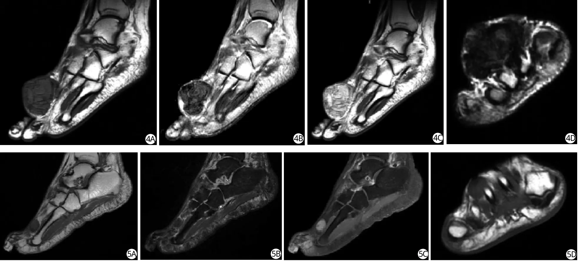
弥漫型腱鞘巨细胞瘤为滑膜样单核细胞的破坏性增生,富含血管,其内还可见多核巨细胞,泡沫细胞、吞噬含铁血黄素的细胞以及炎症细胞;其间质内含有胶原纤维,并常有不同程度的玻璃样变性;在肉眼或镜下它至少局灶性地呈浸润性表现,没有明显边界,具有相当的侵袭性,邻近骨可受侵犯,需广泛切除;可被认为是色素沉着绒毛结节性滑膜炎在关节外软组织内的副本[8-9]。弥漫型腱鞘巨细胞瘤多见于20~50岁之间的年龄段,临床多表现为生长缓慢的质硬、无痛肿块[8]。它是足部最常见的肿物之一,尤其好发于足趾部[4]。在MRI上它多表现为分叶状软组织肿块,部分边界可不清,在T2WI上很大部分的信号不高于肌肉,并可见在T1WI和T2WI上与肌肉相比均呈低信号的区域,增强扫描病灶显著强化[4,9](图4)。局限型腱鞘巨细胞瘤又被称为局限结节性腱鞘滑膜炎,一般可见纤维包膜;发生在关节外,多见于手或足部,尤其是前者;在MRI上其病灶较小,信号较均匀,且边界清楚,邻近骨的髓内侵犯比较少见[10-11](图5)。

图4 足部弥漫型腱鞘巨细胞瘤。矢状面T1WI (A)、T2WI (B) 、增强后T1WI (C)及冠状T2WI (D)示足前部肿物;与肌肉相比病变在T1WI和T2WI呈等、低信号,增强后病变明显强化,邻近第2跖骨被侵蚀破坏 图5 足部局限型腱鞘巨细胞瘤。矢状T1WI (A)、脂肪抑制T2WI (B)、增强后脂肪抑制T1WI (C)及冠状面T1WI (D)示足前部小肿物;与肌肉相比病变在T1WI呈等信号,在T2WI呈低信号,增强后病变明显强化,邻近跖骨未见骨质破坏Fig. 4 Diffuse-type giant cell tumor of tendon sheath in the foot. Sagittal T1-weighted (A),sagital T2-weighted (B), sagittal contrast-enhanced (C), and coronal T2-weighted (D) MR images show a mass in the forefoot with hypo- to isointensity relative to skeletal muscle on both T1- and T2-weighted MR images, marked enhancement and erosion of adjacent second metatarsal bone.Fig. 5 Localized-type giant cell tumor of tendon sheath in the foot. Sagittal T1-wighted (A), sagital fat suppressed T2-weighted(B), sagittal contrast-enhanced (C), and coronal T1-weighted (D) MR images show a small mass in the forefoot with isointensity relative to skeletal muscle on T1-weighted MR image, hypointensity on T2-weighted MR image and marked enhancement.Adjacent metatarsal bone is not.eroded.
5 滑膜肉瘤
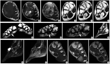
滑膜肉瘤是未分化间叶细胞发生的具有滑膜分化特点的恶性肿瘤;一般发生在关节旁,很少发生于关节腔内;多见于青壮年,男多于女[12]。病灶多贴近或侵犯邻近骨组织,其内常有出血、坏死、钙化,故在T1WI上与肌肉相比以及在T2WI上与脂肪相比常可见高、等、低信号, 注射对比剂后多呈不均匀、显著强化[12](图6)。滑膜肉瘤并非总是信号明显不均,较小的病变(直径不超过5 cm的病变)相对较大病变的信号多较均匀,这在T1WI上表现得尤其明显,而且边界清楚,周围无明显水肿,类似良性病变(图7),易被误诊[12]。
6 Morton神经瘤
Morton神经瘤为足底趾神经的退变及其周围纤维化形成的肿物,可伴有局部血管增生;常发生在3、4跖骨头之间,其次是2、3跖骨间[13-14]。本病好发于女性,表现为前足烧灼样疼痛,放射到第一趾,行走时加重,休息后可缓解[4,14]。其病因可能为跖骨头和跖骨间横韧带对趾神经的压迫引起继发的神经周围纤维化,缺血也可能与其发生有关[14]。MRI上可见病变位于跖骨之间,在跖骨间横韧带的跖侧,以神经血管束为中心;边界清楚;其信号强度在T1WI与肌肉相仿,在T2WI低于皮下脂肪(图8),增强扫描常可见较明显强化;病灶近侧或背侧的跖骨间滑囊可能有积液[13-14]。
7 腱鞘囊肿
腱鞘囊肿是内含黏液的囊性病变,囊壁为纤维结缔组织,没有滑膜内衬,可能为肌腱、韧带、腱鞘、关节囊等纤维组织的黏液样退变所致[4,15]。腱鞘囊肿最好发于手和腕部,足部是其第三好发部位,且尤以足背部多见;它多以可触及的肿块为惟一症状,少数出现疼痛不适、活动受限以及神经卡压等表现[4,15]。在MRI上腱鞘囊肿表现为边界清楚的囊性病变,与肌肉相比在T1WI上均呈稍低信号,在T2WI上呈明显高信号,信号均匀;其内多有分隔,边缘多呈分叶状;增强扫描表现为薄壁强化,囊壁光滑[15](图9)。

图6 足部较大滑膜肉瘤。A:冠状面T1WI示足内侧部肿物,与肌肉相比含高等低三重信号。第一跖骨被破坏。B:冠状面T2WI示肿物与皮下脂肪相比也含高等低三重信号。C:冠状面增强T1WI示增强后肿物呈不均匀、明显强化图7 足部较小滑膜肉瘤。冠状面T1WI (A)、T2WI (B)及增强后T1WI (C)示足底部肿物,边界清楚,与肌肉相比在T1WI呈稍低信号,在T2WI呈高信号,增强后明显强化,其信号较均匀 图8 Morton神经瘤。A:冠状面T1WI示足底第二、三跖骨间小肿物(箭),与肌肉相比呈等信号。B:冠状面T2WI示病变信号稍高于肌肉,低于皮下脂肪。C:冠状面增强T1WI示病灶明显强化 图9 足部腱鞘囊肿。A:矢状面T1WI示足背内侧部肿物,边缘分叶,与肌肉相比呈低信号。B:矢状脂肪抑制T2WI示病变呈明显高信号,内有低信号分隔。C:矢状面脂肪抑制增强T1WI示病变边缘及分隔强化图10 足部表皮样囊肿。A:冠状面T1WI示足底部皮下肿物,无明显分叶,与肌肉相比呈等信号。B:冠状面T2WI示病变主要呈高信号,并含有少量斑片状与肌肉信号相仿的区域。C:冠状增强T1WI显示病变边缘明显强化Fig. 6 Synovial sarcoma with large size in the foot. A: Coronal T1-weighted MR image show a mass in the medial aspect of the foot with hypo, iso and hyperintensity relative to skeletal muscle. The first metatarsal bone is invaded. B: On coronal T2-weighted MR image, the mass show hypo, iso and hyperintensity relative to subcutaneous fat. C: On coronal contrast-enhanced MR image, the mass show heterogeneous and marked enhancement. Fig. 7 Synovial sarcoma with small size in the foot. Coronal T1-weighted (A), T2-weighted (B), and contrast-enhanced (C) MR images show a well de fined mass at the plantar aspect of the foot with relatively homogeneous signal intensity. The mass shows isointensity relative to skeletal muscle on T1-weighted MR image,hyperintensity on T2-weighted MR image and marked enhancement. Fig. 8 Morton neuroma. A: Coronal T1-weighted MR image shows a small mass (arrows) in the plantar aspect of second intermetatarsal space with isointensity relative to skeletal muscle.B: On coronal T2-weighted MR image, the signal intensity of the lesion is slightly higher than that of muscle and lower than that of subcutaneous fat. C: On coronal contrast-enhanced MR image, the lesion shows marked enhancement. Fig. 9 Ganghion cyst in the foot. A: Sagittal T1-weighted MR image shows a lobulated mass in the medial and dorsal aspect of the foot, which demonstrates hypointensity relative to skeletal muscle. B: Sagittal T2-weighted MR image with fat supression shows hyperintense mass with hypointense septa. C: On sagittal contrast-enhanced MR image with fat suppression, the rim and septa of the mass show enhancement. Fig. 10 Epidermoid cyst in the foot. A: Coronal T1- weighted MR image shows a mass in the plantar subcutaneous fat without prominent lobulation. The lesion demonstrates isointensity relative to skeletal muscle. B: On sagittal T2-weighted MR image with fat supression, the mass is mostly hyperintense with a few patchy areas approximately isointense to skeletal muscle. C:On coronal contrast-enhanced MR image with fat suppression, the rim of the mass shows marked enhancement.
8 表皮样囊肿
表皮样囊肿是由皮肤中的表皮细胞在局限的真皮空间内增生所致;组织学上表现为内衬多层鳞状上皮的囊性病变[16-17]。足部的表皮样囊肿通常位于第一跖骨头的足底面或内侧面[2]。MRI检查显示病变位于皮下,多为单房病变,呈圆形或椭圆形,少分叶;与肌肉相比其在T1WI呈等或稍高信号,在T2WI呈高信号;但病变信号可不均匀,其内还常见T2WI信号相对较低的成分,T1WI高信号成分有时也可见,这可能与病变组织学成分不均一,含有角蛋白和胆固醇等成分有关;增强扫描病灶囊壁可强化或不强化[16-17](图10)。
9 小结
根据MRI信号表现可将足部常见软组织肿物分为三类:(1) T2WI信号低于皮下脂肪或含有明显等或低于肌肉信号的肿物,包括纤维性病变、腱鞘巨细胞瘤和Morton神经瘤;前者中的跖腱膜纤维瘤病较小,多位于足底腱膜内侧浅表部位;侵袭性纤维瘤病常累及多块肌肉,一般不侵犯邻近骨的髓腔,在T2WI多以高信号成分为主,混杂带状或条片状较低信号区;弥漫型腱鞘巨细胞瘤侵犯邻近骨的髓腔相对较常见,在T2WI上很大部分的信号不高于肌肉;局限型腱鞘巨细胞瘤较小而局限,边界清楚,少见邻近骨侵犯;Morton神经瘤发病部位较有特点,位于第三、四或二、三跖骨头间、跖骨间横韧带的跖侧,而且好发于女性。(2) T2WI主要呈高信号而低信号成分相对不著的肿物包括血管瘤和滑膜肉瘤;前者在T2WI主要呈明显高信号,其内还常可见血管、脂肪信号,平片中可见静脉石;滑膜肉瘤出血、坏死、囊变较多,在T1WI与肌肉相比以及在T2WI和皮下脂肪相比都可见多重信号,并常贴近或侵犯邻近骨质结构。(3)囊性病变包括腱鞘囊肿和表皮样囊肿;前者常为多房、分叶状,多位于足背内侧;后者多为单房,呈类圆形或椭圆形,少分叶,位于皮下脂肪层,T1WI信号与肌肉相仿或稍高,T2WI虽主要呈高信号,但常可见相对较低信号的成分。
[References]
[1] Bos GD, Esther RJ, Woll TS. Foot tumors: diagnosis and Treatment. J Am Acad Orthop Surg, 2002, 10(4): 259-270.
[2] Llauger J, Palmer J, Monill JM, et al. MR imaging of benign soft-tissue masses of the foot and ankle.Radiographics, 1998, 18(6): 1481-1498.
[3] Teo EL, Strouse PJ, Hernandez RJ. MR imaging differentiation of soft-tissue hemangiomas from malignant soft-tissue masses. AJR Am J Roentgenol, 2000, 174(6):1623-1628.
[4] Waldt S, Rechl H, Rummeny EJ, et al. Imaging of benign and malignant soft tissue masses of the foot. Eur Radiol,2003, 13(5): 1125-1136.
[5] Murphey MD, Ruble CM, Tyszko SM, et al. From the archives of the AFIP: musculoskeletal fibromatoses.Radiologic-pathologic correlation. Radiographics, 2009,29(7): 2143-2173.
[6] Feng ST, Meng QF, Fan M, et al. MRI diagnosis of aggressive fibromatosis. J Clin Radiol, 2005, 25(4):346-349.冯仕庭, 孟悛非, 范淼, 等. 侵蚀性纤维瘤病的MRI诊断.临床放射学杂志, 2005, 25(4): 346-349.
[7] Castellazzi G, Vanel D, Le Cesne A, et al. Can the MRI signal of aggressive fibromatosis be used to predict its behavior? Eur J Radiol, 2009, 69(2): 222-229.
[8] Somerhausen NS, Fletcher CD. Diffuse-type giant cell tumor: clinicopathologic and immunohistochemical analysis of 50 cases with extraarticular disease. Am J Surg Pathol, 2000, 24(4): 479-492.
[9] Murphey MD, Rhee JH, Lewis RB, et al. Pigmented villonodular synovitis: radiologic-pathologic correlation.Radiographics, 2008, 28(5): 1493-518.
[10] Monaghan H, Salter DM, Al-Nafussi A. Giant cell tumour of tendon sheath (localized nodular tenosynovitis):clinicopathological features of 71 cases. J Clin Pathol,2001, 54(5): 404-407.
[11] Kitagawa Y, Ito H, Amano Y, et al. MR imaging for preoperative diagnosis and assessment of local tumor extent on localized giant cell tumor of tendon sheath.Skeletal Radiol, 2003, 32(11): 633-638.
[12] Zhang ZH, Meng QF, Zhang XL. MRI diagnosis of synovial sarcoma of extremities. J Clin Radiol, 2006,25(10): 941-944.张朝晖, 孟悛非, 张小玲. 滑膜肉瘤的MRI诊断. 临床放射学杂志, 2006, 25(10):941-944.
[13] Abreu E, Aubert S, Wavreille G, et al. Peripheral tumor and tumor-like neurogenic lesions. Eur J Radiol, 2011[Epub ahead of print]DOI:10.1016/j.ejrad.2011.04.036.
[14] Ashman CJ, Klecker RJ, Yu JS. Forefoot pain involving the metatarsal region: differential diagnosis with MR imaging. Radiographics, 2001, 21(6): 1425-1440.
[15] Zhang ZH, Liang MQ, Li ZH. MRI diagnosis of the soft tissue ganglion cyst in the foot and ankle. Diagnostic imaging & Interventional Radiology, 2011, 20(6): 52-54.张朝晖, 梁满球, 李竹浩. 足踝部软组织腱鞘囊肿的MRI诊断. 影像诊断与介入放射学杂志, 2011, 20(6): 52-54.
[16] Hong SH, Chung HW, Choi JY, et al. MRI findings of subcutaneous epidermal cysts: emphasis on the presence of rupture. AJR Am J Roentgenol, 2006, 186(4): 961-966.
[17] Shibata T, Hatori M, Satoh T, et al. Magnetic resonance imaging features of epidermoid cyst in the extremities.Arch Orthop Trauma Surg, 2003, 123(5): 239-241.
