Pre-ischemia electro-acupuncture potentiates the expression of Bcl-2 and transforming growth factor-beta 1 in rat brains*☆△◇
2012-09-12KaKeungYipSamuelCLLoKwokfaiSoDoraMYPoonMasonCPLeung
Ka Keung Yip, Samuel CL Lo, Kwok-fai So, Dora MY Poon, Mason CP Leung
1 Department of Rehabilitation Sciences, the Hong Kong Polytechnic University, Hung Hom, Hong Kong Special Administrative Region, China
2 Department of Applied Biology and Chemical Technology, the Hong Kong Polytechnic University, Hung Hom, Hong Kong Special Administrative Region, China
3 Department of Anatomy, the University of Hong Kong, Pokfulam, Hong Kong Special Administrative Region, China
Pre-ischemia electro-acupuncture potentiates the expression of Bcl-2 and transforming growth factor-beta 1 in rat brains*☆△◇
Ka Keung Yip1, Samuel CL Lo2, Kwok-fai So3, Dora MY Poon1, Mason CP Leung1
1 Department of Rehabilitation Sciences, the Hong Kong Polytechnic University, Hung Hom, Hong Kong Special Administrative Region, China
2 Department of Applied Biology and Chemical Technology, the Hong Kong Polytechnic University, Hung Hom, Hong Kong Special Administrative Region, China
3 Department of Anatomy, the University of Hong Kong, Pokfulam, Hong Kong Special Administrative Region, China
Abstract
The expression of the anti-apoptotic molecules Bcl-2 and transforming growth factor-beta 1 is known to confer protective effects on the cerebral ischemia-reperfusion injury. The current study investigated the expression levels of Bcl-2 and transforming growth factor-beta 1 in response to multiple pre-ischemia electro-acupuncture at acupoints Zusanli (ST36) and Fengchi (GB20)stimulation. Rats were divided into five groups: uninjured, control, non-acupoint, GB20 and ST36.Rats in the non-acupoint, GB20 and ST36 groups received 30 minutes (3 times or 18 times) of electro-acupuncture stimulation before experimental cerebral ischemia was induced. Bcl-2 and transforming growth factor-beta 1 were found to be significantly increased in the ST36 groups with either 3 or 18 electro-acupuncture treatments (P < 0.05). The production was higher with 18 electro-acupuncture treatments in the ST36 groups (P < 0.05). In the GB20 groups, significant increase was only observed in transforming growth factor-beta 1 with 18 electro-acupuncture treatments (P < 0.05). No significant elevation of the level of transforming growth factor-beta 1 was observed in the non-acupoint groups. However, the production of Bcl-2 increased with 18 treatments in the non-acupoint groups (P < 0.05). The data suggest that multiple pre-ischemia electro-acupuncture at ST36 was effective in conferring neuroprotective effect on the brain by means of upregulation of Bcl-2 and transforming growth factor-beta 1 and the effect was increase with the number of treatment.
Key Words
cerebral ischemia; stroke prevention; electro-acupuncture; transforming growth factor-beta 1; Bcl-2;acupoint
Research Highlights
(1) With the success of post-ischemia application of electro-acupuncture, scientists start their interest in its preventive ability.
(2) Two groups of multiple pre-ischemia electro-acupuncture treatment, 3 times and 18 times, were applied bilaterally at Zusanli (ST36), Fengchi (GB20) and non-acupoint.
(3) Beneficial effect which favored more session of treatment was observed only at ST36 after cerebral ischemia through up-regulating the level of Bcl-2 and transforming growth factor-beta 1.
Ka Keung Yip☆, MPhil,
Research Associate,Department of Rehabilitation Sciences, the Hong Kong Polytechnic University, Hung Hom, Hong Kong, Special Administrative Region, China
Corresponding author:Mason CP Leung, Assistant professor, Department of Rehabilitation Sciences, the Hong Kong Polytechnic University, Hung Hom, Hong Kong Special Administrative Region, China
rsdpoon@polyu.edu.hk
Received: 2012-05-21 Accepted: 2012-07-06(NY20120530001/ZLJ)
Yip KK, Lo SCL, So KF, Poon DMY, Leung MCP.
Pre-ischemia electro-acupuncture potentiates the expression of Bcl-2 and transforming growth factor-beta 1 in rat brains. Neural Regen Res.2012;7(24):1859-1865.
www.crter.cn
www.nrronline.org
INTRODUCTION
Transforming growth factor-beta 1 is an ubiquitous cytokine that influences various cell types, including microglia[1]and neurons[1-2]. It is virtually absent from intact brain tissue[2-3], but its expression increases strongly after trauma[2], excitotoxic lesioning1 and ischemia-reperfusion injury[4]. Transforming growth factor-beta 1 induction has been associated with several anti-inflammatory effects including decreased neuronal susceptibility to glutamate excitotoxicity, macrophage deactivation and astrocytic brain-derived neurotrophic factor induction[3]. transforming growth factor-beta 1 also contributes to calcium homeostasis in nerve cells, which is important for the regulation of neuronal nitric oxide synthase expression[1]. Transforming growth factor-beta 1 also reduces cerebral ischemia-reperfusion induced inducible nitric oxide synthase expression[5]. Increased transforming growth factor-beta 1 protein levels may act as an initiator of neuroprotective mechanisms. Bcl-2 is a known oncogene product that protects cells from apoptosis by preventing the release of cytochrome C[6-7].Increased Bcl-2 expression is also neuroprotective because it decreases the formation of peroxynitrite and nitric oxide, thereby reducing infarct volume and inhibiting the activation of caspase-3[8-10].Acupuncture, in particular electro-acupuncture, has increasingly been used as a complementary therapy for pain relief[11-12]and stroke rehabilitation in both Asian and western countries[13-15]. A considerable number of studies investigated the effectiveness of electro-acupuncture therapy on patients with cerebral ischemia. Several beneficial outcomes have been noted, including reduced paralysis due to increased muscle strength[16-17],improved speech[17], reduced mental retardation[17],restored cerebral blood flow[18-19]and improved locomotion[17-18,20-21]. These observations support the hypothesis that post-ischemia electro-acupuncture can be an effective modality for the treatment of stroke.However, little is known about the effectiveness of pre-ischemia electro-acupuncture in minimizing neurological injury caused by stroke. If proven successful,pre-ischemia electro-acupuncture could be used as a preventive measure for patients at increased risk of stroke. In this study, we investigated the effects of multiple applications of electro-acupuncture stimulation before the induction of cerebral ischemia. Two acupoints,Fengchi (GB20) and Zusanli (ST36), were selected for this study because electro-acupuncture stimulation of these two points might minimize damage to brain tissue through its ability to attenuate lipid peroxidation in areas affected by stroke[22]. Furthermore, GB20 is an effective acupoint often selected for stroke rehabilitation because treatment via GB20 greatly improves the locomotive ability of stroke patients[18,21]. In contrast, ST36 is a common acupoint for analgesia, spasmolysis and homeostasis[23-24].
In order to confirm the correct selection of acupoint in the tissue, the bilateral non-acupoint was selected and compared to the selected acupoint. After the induction of cerebral ischemia, the effects of electro-acupuncture stimulation at different acupoints were assessed by measuring the expression of transforming growth factor-beta 1 and Bcl-2. As described above, the increased production of transforming growth factor-beta 1 and Bcl-2 has been shown to be neuroprotective against cerebral ischemia.
RESULTS
Physiological parameters
The physiological data obtained before and during
transient focal cerebral ischemia are shown in Table 1.

Table 1 Physiological data from rats subjected to transient focal cerebral ischemia
The values of the parameters measured were consistent with previously reported values[22,25]. The difference between the values of partial oxygen pressure measured before and after ischemia was not statistically significant(t = 0.846, degrees of freedom (d.f.) = 5, a = 0.05).Similarly, the values of partial carbon dioxide pressure measured before ischemia, was comparable to that after middle cerebral artery occlusion (t = 0.270, d.f. = 5, a =0.05). In addition, there was no significant difference between the values of blood pH measured before and after cerebral ischemia. These findings suggest that the observed changes in cerebral tissues were not a result of physiological changes.
Expression of transforming growth factor-beta 1 in the ischemic hemisphere of the brain
The expression of transforming growth factor-beta 1 in the different groups is shown in Figure 1. Transforming growth factor-beta 1 was not detected in the uninjured group. After three applications of electro-acupuncture stimulation, no significant differences were observed between the control, non-acupoint and GB20 groups.
However, transforming growth factor-beta 1 expression was significantly higher in the ST36 group when compared to the other three groups (P < 0.05). Similarly,after 18 applications of electro-acupuncture stimulation,the highest level of transforming growth factor-beta 1 was detected in the ST36 group. This expression level was significantly higher than those of the other three groups (P < 0.05). In addition, the GB20 group with the same treatment frequency also produced two-fold more transforming growth factor-beta 1 than the control group(P < 0.05). Nevertheless, an increase in transforming growth factor-beta 1 expression associated with a greater number of treatments was observed in the ST36 group in which 18 electro-acupuncture applications induced about 50% more transforming growth factor-beta 1 than did 3 electro-acupuncture applications (P < 0.05).

Figure 1 Effect of pre-ischemia electro-acupuncture on expression of transforming growth factor-beta 1 (TGF-β1)in the ischemic hemisphere of the brain.
Expression of Bcl-2 in the ischemic hemisphere of the brain
The expression of Bcl-2 in the different groups is shown in Figure 2. The amount of Bcl-2 expressed in the uninjured group was 0.502 ± 0.06 pg Bcl-2 per mg protein. With three applications of electro-acupuncture stimulation, a significant increase in Bcl-2 expression was only observed in the ST36 group (P < 0.05). Bcl-2 expression was higher with 18 applications of electro-acupuncture stimulation than with 3 applications in all groups. Relative to the control and GB20 groups,the ST36 group expressed 29% and 15% (respectively)more Bcl-2 (P < 0.05). However, Bcl-2 expression was also upregulated in the non-acupoint group (18-times),similar to the expression in the ST36 group.
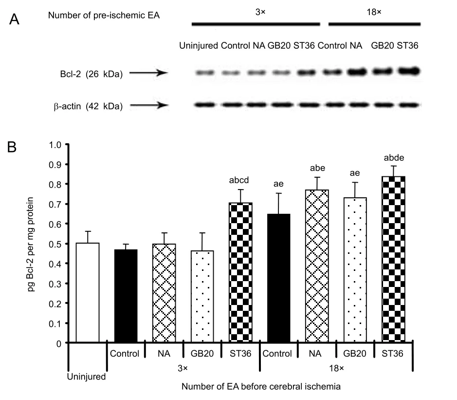
Figure 2 Effect of pre-ischemia electro-acupuncture on expression of Bcl-2 in the ischemic hemisphere of the brain.
DISCUSSION
The current study investigated the effect of pre-ischemia electro-acupuncture on the expression of transforming
growth factor-beta 1 and Bcl-2 at day 4 post-injury. It has been previously reported that transforming growth
factor-beta 1 mRNA expression increases after transientand permanent occlusion of middle cerebral artery[3,26].In the present study, the expression of transforming growth factor-beta 1 was not detected in the uninjured group. Similar findings were also reported in the expression of transforming growth factor-beta 1 mRNA in unoperated rats[1]. After a 1-hour occlusion of middle cerebral artery, a significant increase in the level of transforming growth factor-beta 1 protein in the ST36 group (3-time electro-acupuncture and 18-time electro-acupuncture) and the GB20 group (18-time electro-acupuncture) was observed. The administration of transforming growth factor-beta 1 has been reported to decrease infarct size after focal cerebral ischemia in rats[27-28]and rabbits[29], and to protect hippocampal neurons after global cerebral ischemia[2]. Therefore,pre-ischemia electro-acupuncture may have a beneficial effect in neuroprotection after cerebral ischemia by upregulating the content of transforming growth factor-beta 1. The neuroprotection may take place in penumbral region of the cerebral cortex and striatum where the elevated expression of transforming growth factor-beta 1 mRNA following occlusion of middle cerebral artery was reported recently[30].
Bcl-2 also plays an important role in neuroprotection in cerebral ischemia. Decreased infarct area was observed in over-expressed Bcl-2 transgenic mice[31], whereas increased infarct area was found in Bcl-2 knockout mice[32]. The neuronal protection controlled by Bcl-2 is through the inhibition of free radical production[33],cytosolic accumulation of cytochrome C and caspase-3 activation[10]. In the current study, after pre-ischemia electro-acupuncture, the level of Bcl-2 was elevated at ST36 (3-times and 18-times) but not at GB20. In contrast of our previous study, pre-ischemia electro-acupuncture at GB20 showed that malondialdehyde production was inhibited[22]suggesting that different acupoints may exert neuronal protection through different biochemical pathways. Hippocampal CA1 region was most frequently employed for Bcl-2 investigation under cerebral ischemic condition due to its vulnerability to cerebral injury.Ferrer et al[34]observed a marked increase of Bcl-2 expression in hippocampal CA1 region at post-injury day 4. Zhang and Wang[35]also reported that Bcl-2 mRNA was up-regulated in hippocampus following cerebral ischemia. In a recent study, electro-acupuncture at ST36 and GV20 were able to increase Bcl-2 expression in hippocampal CA1 area[36].
In both production profiles of transforming growth factor-beta 1 and Bcl-2, the up-regulation was higher in 18-time application when compared with those after 3 times at pre-ischemia electro-acupuncture at ST36. More benefits might be derived from a higher number of pre-conditioning electro-acupuncture sessions. However,the current study employed only two time points, which might not be able to determine if and when any plateau effect or adverse effect may be achieved.
Apart from the current beneficial effect of pre-ischemia electro-acupuncture on cerebral ischemia, previous studies also revealed that reduced infarct size,neurological deficit and apoptosis were observed even in a single 30-minutes pre-ischemia electro-acupuncture at acupoint GB20 through the regulation of endocannabinoid system[37]. Dong et al[38]also demonstrated that repeated electro-acupuncture preconditioning (5 days) at acupoint GB20 could lower the infarct size and neurological deficit score with the inhibition of matrix metalloproteinase-9 expression and activity. Our research team also reported in a previous study that multiple pre-ischemia electro-acupuncture at ST36 and GB20 could reduce the production of malonadialdehyde[22].
Although previous studies have focused on stroke rehabilitation by electro-acupuncture stimulation[13-15], the present study explored another clinical application of electro-acupuncture stimulation. Pre-ischemia electro-acupuncture stimulation is particularly important for specific high-risk groups, such as patients with high blood pressure or other congenital characteristics that predispose them to stroke[39]. Applying electro-acupuncture stimulation at either GB20 or ST36 on alternate days each week might ameliorate the severity of brain damage after a stroke and shorten the time of recovery.
Data from this study confirmed that multiple pre-injury electro-acupuncture stimulations at ST36 could effectively increase the production of both transforming growth factor-beta 1 and Bcl-2, with an up-regulation of transforming growth factor-beta 1 correlated to a neuroprotective effect at GB20. These proteins work together to reduce the extent of brain tissue damage after cerebral ischemia-reperfusion. Electro-acupuncture treatment can be used as a preventive measure against stroke damage. In addition, transforming growth factor-beta 1 promotes the neuroprotection of proteins against expected oxidative stress by regulating the apoptotic cascade. The small sample size of this study is an obvious limitation; further experiments with a larger number of animals are required for a more detailed evaluation of apoptotic events.
MATERIALS AND METHODS
Design
A randomized controlled animal experiment.
Time and setting
This experiment was performed at the Hong KongPolytechnic University, Hong Kong Special Administrative Region, China from 2007 to 2009.
Materials
Animals
All experimental procedures were approved by the Hong Kong Polytechnic University animal ethical committee. A total of 68 male Sprague-Dawley rats weighing 300-350 g were used. Firstly, 12 rats were used to measure common physiological parameters in blood obtained from the femoral artery 15 minutes before and during cerebral ischemia. Then eight rats were assigned to the uninjured group for measurement of the parameters listed below. Forty-eight rats were evenly divided into four experimental groups: a control group (rats with general anesthesia without electro-acupuncture), a non-acupoint group (rats with electro-acupuncture stimulation at non-acupoint), a GB20 group (rats with electro-acupuncture stimulation at GB20), and an ST36 group (rats with electro-acupuncture stimulation at ST36).electro-acupuncture stimulation was carried out on alternate days before the induction of cerebral ischemia.In each group, six rats were subjected to 3 electro-acupuncture treatments conducted for a week and another six rats were subjected to 18 electro-acupuncture treatments conducted over 6 consecutive weeks. Twenty-four hours after the last electro-acupuncture treatment, the rats in the experimental groups were then subjected to transient focal cerebral ischemia followed by harvesting of their brains on post-ischemia day 4. The sampling point was determined by considering the maximum nitric oxide synthase activity after the induction of cerebral ischemia[40].
Chemicals and antibodies
Unless otherwise stated, all materials were of analytical grade and obtained from Sigma Chemical Co. (St. Louis,MO, USA). Primary antibodies (200 μg/mL) against transforming growth factor-beta 1 and Bcl-2 were used at a dilution of 1:200. β-actin was employed as an internal control at a dilution of 1:2 000. Secondary antibodies conjugated with horseradish peroxidase directed against rabbit IgG were obtained from Pierce Chemical Co. (Rockford, IL, USA) and used at a dilution of 1:1 000. Protein concentrations were determined with a protein assay (Bio-Rad Laboratories, Hercules, CA,USA).
Methods
Pre-ischemia electro-acupuncture
A 30-minute electro-acupuncture treatment was applied by an acupunctoscope device (Model G6805-2, Smeif,Shanghai, China) (Voltage: 0.7 V, frequency: 2 Hz,duration: 0.5 ms) as previously described[22]. In the GB20 and ST36 groups, electro-acupuncture was applied at corresponding bilateral points. GB20 is anatomically located on the posterior aspect of the neck, below the occipital bone, in the depression between the sternocleidomastoid muscle and the trapezius muscle[41].ST36 is anatomically located near the knee joint of the hind limb 2 mm lateral to the anterior tubercle of the tibia[41]. In the non-acupoint group, electro-acupuncture was applied at a non-acupoint located midway between the coccyx and the hip joint. The results from the use of non-acupoints for control purposes highlight the significance of selecting the correct acupoint. Apart from electrical stimulation, needle puncture is also one of the acupoint stimulation methods, so it is not appropriate to serve as a control. Previous studies reported that treatment of acupuncture at ST36 could alleviate ischemia-induced apoptosis[42], inhibit inflammation[43]and promote neurogenesis[44].
Induction of transient focal cerebral ischemia
Transient focal cerebral ischemia was induced by the occlusion of the right middle cerebral artery for 1 hour.Rats were anesthetized by an intraperitoneal injection of ketamine (70 mg/kg, Alfasan, Holland) and xylazine(7 mg/kg, Alfasan, Holland). The temporalis muscle was then briefly separated in the plane of its fiber bundles to expose the zygoma and squamosal bones. A 5-mm x 5-mm burr hole was made to expose the middle cerebral artery, which was then occluded for 1 hour followed by reperfusion. During the surgical procedure, the rectal temperature of the rats was monitored and maintained at about 37°C with an overhead lamp. After recovery from the experimental surgery, all rats were allowed to recover at ambient temperature (21-23°C) with food and water made available ad libitum until harvest. To measure physiological parameters, arterial blood samples were taken from the femoral artery 15 minutes before and after middle cerebral artery occlusion for the determination of pH, arterial partial pressure of oxygen and carbon dioxide by a Blood Gas and Electrolyte System
(Radiometer ABL505, Copenhagen, Denmark).
Sample collection
Rats were sacrificed 4 days after ischemia-reperfusion with an overdose ketamine and xylazine mixture.
Transcardial perfusion was performed with 0.38%sodium citrate in normal saline for 10 minutes followed by 0.9% sodium chloride for 5 minutes. The harvested right hemisphere of the brain was weighted and homogenized in 20 mM Tris-HCl (pH 7.4). The homogenate was centrifuged at 3 000 × g for 10 minutes at 4°C and the supernatant was collected for analysis.
Determination of transforming growth factor-beta 1 and Bcl-2 expression
Transforming growth factor-beta 1 and Bcl-2 were quantitated by western blots probed with specific primary antibodies. A 15% SDS-PAGE resolving gel was prepared for western blotting for the detection of transforming growth factor-beta 1 and Bcl-2. In brief,protein homogenate (200 μg) resolved by gel electrophoresis was electroblotted to nitrocellulose membranes at a constant voltage of 25 V for 16 hours.After transfer of the proteins, the membranes were blocked with skim milk in 1× TBS-T, and they were then probed with primary antibodies raised against transforming growth factor-beta 1 and Bcl-2 overnight at 4°C with gentle shaking. Subsequently, the probed blots were incubated with secondary antibodies with gentle shaking. The immunolabeled proteins were detected by their reaction with a chemiluminescent substrate (Pierce Chemical Co., Rockford, IL, USA). The chemiluminescence released was captured and quantified using known amount of loaded analytes as reference. Selected blots were re-probed with β-actin as an internal control.
Statistical analysis
Data were expressed as the mean ± SD. Statistical analysis of the physiological parameters was performed using the paired t-test (SPSS 14.0 software,SPSS, Chicago, IL, USA). Multivariate analysis of the other parameters showed a significant interaction between two factors: the selected acupoint and the number of pre-ischemic electro-acupuncture stimulation treatments. Therefore, comparisons were made among these groups using the analysis of variance method followed by the post-hoc protected least-significant difference test. In all cases, P < 0.05 is considered to be statistically significant.
Acknowledgments: The authors would like to thank Ms.Christine Van for editing.
Funding: This study was supported by the Niche Area Grant of the Hong Kong Polytechnic University through the projects JBB71 and BB8V.
Author contributions: Ka Keung Yip was responsible for manuscript writing and obtaining experimental data. Samuel C.L. Lo, Kwok-fai So and Dora M.Y. Poon participated in study design and technical instruction. Mason C.P. Leung was in charge of study assessment, manuscript authorization and the funding process.
Conflicts of interest: None declared.
Ethical approval: The study was approved by the Animal
Subjects Ethics Sub-Committee of The Hong Kong Polytechnic University (ASESC 06/6), China.
REFERENCES
[1] Lehrmann E, Kiefer R, Finsen B, et al. Cytokines in cerebral ischemia: expression of transforming growth factor beta-1 (TGF-beta 1) mRNA in the postischemic adult rat hippocampus. Exp Neurol. 1995;131(1):114-123.
[2] Henrich-Noack P, Prehn JH, Krieglstein J. TGF-beta 1 protects hippocampal neurons against degeneration caused by transient global ischemia: dose-response relationship and potential neuroprotective mechanisms.Stroke. 1996;27(9):1609-1614.
[3] Lehrmann E, Kiefer R, Christensen T, et al. Microglia and macrophages are major sources of locally produced transforming growth factor-beta1 after transient middle cerebral artery occlusion in rats. Glia. 1998;24(4):437-448.
[4] Wang J, Wang L. Clinical observation on 186 cases of apoplexy treated with acupuncture. Zhongguo Zhen Jiu 1995;15:11-12.
[5] Thomas AG, Liu W, Olkowski JL, et al. Neuroprotection mediated by glutamate carboxypeptidase II (NAALADase)inhibition requires TGF-beta. Eur J Pharmacol. 2001;430(1):33-40.
[6] Knox KA, Johnson GD, Gordon J. A study of protein kinase C isozyme distribution in relation to Bcl-2 expression during the apoptosis of epithelial cells in vivo.Exp Cell Res. 1993;207(1):68-73.
[7] Ojala PM, Yamamoto K, Castaños-Vélez E, et al. The apoptotic v-cyclin-CDK6 complex phosphorylates and inactivates Bcl-2. Nat Cell Biol. 2000;2(11):819-825.
[8] Cao G, Luo Y, Nagayama T, et al. Cloning and characterization of rat caspase-9: implications for a role in mediating caspase-3 activation and hippocampal cell death after transient cerebral ischemia. J Cereb Blood Flow Metab. 2002;22(5):534-546.
[9] Seo JH, Rah JC, Choi SH, et al. Alpha-synuclein regulates neuronal survival via Bcl-2 family expression and PI3/Akt kinase pathway. FASEB J. 2002;16(13):1826-1828.
[10] Zhao H, Yenari MA, Cheng D, et al. Bcl-2 overexpression protects against neuron loss within the ischemic margin following experimental stroke and inhibits cytochrome c translocation and caspase-3 activity. J Neurochem.2003;85(4):1026-1036.
[11] Romita VV, Suk A, Henry JL. Parametric studies on electroacupuncture-like stimulation in a rat model: effects of intensity, frequency, and duration of stimulation on evoked antinociception. Brain Res Bull. 1997;42(4):289-296.
[12] Tsui P, Leung MCP. Comparison of the effectiveness between manual acupuncture and electro-acupuncture on patients with tennis elbow. Acupunct Electrother Res.2002;27(2):107-117.
[13] Cheng X. Chinese Acupuncture and Moxibustion. Beijing:Foreign Language Press. 1987.
[14] Leake R, Broderick JE. Treatment efficacy of acupuncture:a review of the research literature. Integr Med. 1998;1:107-115.
[15] Si QM, Wu GC, Cao XD. Effects of electroacupuncture on acute cerebral infarction. Acupunct Electrother Res. 1998;23(2):117-124.
[16] Huang WD, Mou XX, Hao XS, et al. Effect of acupoints in heart meridian and Neiguan on cardiac function of patients with ischemic heart disease. Zhongguo Zhen Jiu.1998;18:494-496.
[17] Xing QZ, Zhang SW. Clinical observation of 75 cases of cerebral haemorrhage treated with acupuncture.Zhongguo Zhen Jiu. 1998;18:719-722.
[18] Tan GL. Clinical observation of 105 cases of occluded cerebral artery treated with acupuncture at Fengchi.Zhongguo Zhen Jiu. 1990;10:21-22.
[19] Zhai N, Du Y, Shi X, et al. Morphological study on acupuncture in interfering experimental cerebral infarction in rat. I. Compensation of cerebral PIA mater artery in cerebral surface. Zhen Ci Yan Jiu. 1993;18(1):8-13.
[20] Chang YH, Hsieh MT, Cheng JT. Increase of locomotor activity by acupuncture on Bai-Hui point in rats. Neurosci Lett. 1996;211(2):121-124.
[21] Liu YZ, Yang JS, Zhang GR, et al. Effect of acupuncture on locomotion of patients after stroke. Zhongguo Zhen Jiu.1999;19:69-71.
[22] Siu FKW, Lo SCL, Leung MCP. Effectiveness of multiple pre-ischemia electro-acupuncture on attenuating lipid peroxidation induced by cerebral ischemia in adult rats.Life Sci. 2004;75(11):1323-1332.
[23] Stux G, Pomeranz B. Basic of Acupuncture. Berlin:Springer. 1988;86-87.
[24] Yun SJ, Park HJ, Yeom MJ, et al. Effect of electroacupuncture on the stress-induced changes in brain-derived neurotrophic factor expression in rat hippocampus. Neurosci Lett. 2002;318(2):85-88.
[25] Leker RR, Teichner A, Ovadia H, et al. Expression of endothelial nitric oxide synthase in the ischemic penumbra: relationship to expression of neuronal nitric oxide synthase and vascular endothelial growth factor.Brain Res. 2001;909(1-2):1-7.
[26] Yamashita K, Gerken U, Vogel P, et al. Biphasic expression of TGF-beta1 mRNA in the rat brain following permanent occlusion of the middle cerebral artery. Brain Res. 1999;836(1-2):139-145.
[27] Prehn JH, Backhauss C, Krieglstein J. Transforming growth factor-beta 1 prevents glutamate neurotoxicity in rat neocortical cultures and protects mouse neocortex from ischemic injury in vivo. J Cereb Blood Flow Metab.1993;13(3):521-525.
[28] McNeill H, Williams C, Guan J, et al. Neuronal rescue with transforming growth factor-beta 1 after hypoxic-ischaemic brain injury. Neuroreport. 1994;5(8):901-904.
[29] Gross CE, Bednar MM, Howard DB, et al. Transforming growth factor-beta 1 reduces infarct size after experimental cerebral ischemia in a rabbit model. Stroke.1993;24(4):558-562.
[30] Vincze C, Pál G, Wappler EA, et al. Distribution of mRNAs encoding transforming growth factors-beta1, -2, and -3 in the intact rat brain and after experimentally induced focal ischemia. J Comp Neurol. 2010;518(18): 3752-3770.
[31] Martinou JC, Dubois-Dauphin M, Staple JK, et al.Overexpression of BCL-2 in transgenic mice protects neurons from naturally occurring cell death and experimental ischemia. Neuron. 1994;13(4):1017-1030.
[32] Hata R, Gillardon F, Michaelidis TM, et al. Targeted disruption of the bcl-2 gene in mice exacerbates focal ischemic brain injury. Metab Brain Dis. 1999;14(2):117-124.
[33] Lee M, Hyun DH, Marshall KA, et al. Effect of overexpression of Bcl-2 on cellular oxidative damage,nitric oxide production, antioxidant defences, and the proteasome. Free Radic Biol Med. 2001;31(12):1550-1559.
[34] Ferrer I, López E, Blanco R, et al. Bcl-2, Bax, and Bcl-x expression in the CA1 area of the hippocampus following transient forebrain ischemia in the adult gerbil. Exp Brain Res. 1998;121(2):167-173.
[35] Zhang S, Wang W. Altered expression of bcl-2 mRNA and Bax in hippocampus with focal cerebral ischemia model in rats. Chin Med J (Engl). 1999;112(7):608-611.
[36] Bu Y, Zhang PY, Geng DQ, et al. Effects of electroacupuncture plus oxygenmedicine on the expression of Bcl-2 and Bax proteins in the hippocampal CA 1 area in rats with global cerebral ischemia/reperfusion injury. Zhen Ci Yan Jiu. 2010;35(3):208-212.
[37] Wang Q, Peng Y, Chen S, et al. Pretreatment with electroacupuncture induces rapid tolerance to focal cerebral ischemia through regulation of endocannabinoid system. Stroke. 2009;40(6):2157-2164.
[38] Dong H, Fan YH, Zhang W, et al. Repeated electroacupuncture preconditioning attenuates matrix metalloproteinase-9 expression and activity after focal cerebral ischemia in rats. Neurol Res. 2009;31(8):853-858.
[39] Harmsen P, Lappas G, Rosengren A, et al. Long-term risk factors for stroke: twenty-eight years of follow-up of 7457 middle-aged men in Göteborg, Sweden. Stroke. 2006;37(7):1663-1667.
[40] Leung MCP, Lo SCL, Siu FKW, et al. Treatment of experimentally induced transient cerebral ischemia with low energy laser inhibits nitric oxide synthase activity and up-regulates the expression of transforming growth factor beta-1. Lasers Surg Med. 2002;31(4):283-288.
[41] Ellis A, Wiseman N, Boss K. Fundamentals of Chinese Acupuncture. Massachusetts: Paradigm Publications.1991.
[42] Chung JH, Lee EY, Jang MH, et al. Acupuncture decreases ischemia-induced apoptosis and cell proliferation in dentate gyrus of gerbils. Neurol Res. 2007;29(Suppl 1):S23-S27.
[43] Chae Y, Hong MS, Kim GH, et al. Protein array analysis of cytokine levels on the action of acupuncture in carrageenan-induced inflammation. Neurol Res. 2007;29(Suppl 1):S55-S58.
[44] Hwang IK, Chung JY, Yoo DY, et al. Comparing the effects of acupuncture and electroacupuncture at Zusanli and Baihui on cell proliferation and neuroblast differentiation in the rat hippocampus. J Vet Med Sci. 2010;72(3):279-284.
(Edited by Gioltzoglou T, Cesca F/Song LP)
10.3969/j.issn.1673-5374.2012.24.003
杂志排行
中国神经再生研究(英文版)的其它文章
- Effects of wind-dispelling drugs and deficiency-nourishing drugs of Houshiheisan compound prescription on astrocyte activation and inflammatory factor expression in the corpus striatum of cerebral ischemia rats****☆
- Acupuncture and moxibustion for visceral pain
- Therapeutic effect of nerve growth factor on cerebral infarction in dogs using the hemisphere anomalous volume ratio of diffusion-weighted magnetic resonance imaging*★
- Yizhijiannao Granule and a combination of its effective monomers, icariin and Panax notoginseng saponins, inhibit early PC12 cell apoptosis induced by beta-amyloid (25-35)☆
- Acupuncture activates signal transduction pathways related to brain-tissue restoration after ischemic injury**☆
- No association between a polymorphism of the adenylate cyclase type IX gene and major depressive disorder in the Chinese Han population*☆