受翻译调节的肿瘤蛋白在PRL-3促进结肠癌转移中的作用*
2011-09-14褚忠华李守峰曾育杰关玉峰
褚忠华, 刘 璐, 来 伟, 李守峰, 曾育杰, 关玉峰
(1中山大学孙逸仙纪念医院胃肠外科,广东广州510120;2广州市番禺中心医院普通外科,广东广州511400)
肝再生磷酸酶-3(phosphatase of regenerating liver-3,PRL-3)是近年来发现的一种具有促进细胞增殖、迁移和侵袭的蛋白酪氨酸磷酸酶[1]。最初,Saha等[2]通过比较结直肠癌转移灶与原发肿瘤之间的基因表达差异,发现PRL-3在结直肠癌肝转移灶中高度表达,同时存在基因拷贝数的增加,而在未转移的结直肠癌和正常的结直肠上皮不表达。之后,陆续有研究发现PRL-3在卵巢癌、胃癌、乳腺癌和霍奇金淋巴瘤中表达升高[3-6]。目前,已有大量研究表明PRL-3在多种肿瘤细胞的转移过程中均起到重要作用[3,4,6]。然而 PRL -3 促进肿瘤转移的具体分子机制仍然不清楚。受翻译调节的肿瘤蛋白(translationally controlled tumor protein,TCTP),是一类广泛存在于动物、植物及酵母中,并在序列上高度保守且有很高同源性的蛋白家族[7]。最初认为TCTP是一类生长相关蛋白,但随后的研究提示TCTP可能具有非常重要的生物学功能,包括调节细胞周期的进程和恶性转移,钙结合蛋白和细胞外组胺释放蛋白等[7]。更重要的是,新近的研究发现,TCTP在前列腺癌和结肠癌细胞增殖、细胞凋亡、细胞迁移和侵袭及转移均具有重要作用[8,9]。由于PRL-3与TCTP在肿瘤转移的多个环节均具有重要调节作用,因此它们在发挥促肿瘤转移过程中可能存在一定的相关性。因此,本文主要探讨PRL-3是否对TCTP的表达进行调节及TCTP在PRL-3促结肠癌转移中的作用。
材料和方法
1 材料
结肠癌细胞株LoVo购自中山大学医学院细胞库,大肠杆菌DH-5α购自上海生工公司,真核载体pAcGFP -C3购自 Clontech,T4连接酶、Xho I、EcoR I内切酶、Trizol和RT-PCR试剂盒购自TaKaRa,Lipofectamine 2000和G418购自Invitrogen。兔抗PRL-3多克隆抗体和兔抗TCTP多克隆抗体购自Santa Cruz,辣根过氧化物酶标记的羊抗兔Ⅱ抗购自Santa Cruz。siRNA干扰序列由上海吉马公司合成,CCK-8试剂盒购自碧云天公司,Transwell双层细胞培养板(直径 6.5 mm,孔径 8.0 μm)购自 Corning,matrigel购自BD,4%多聚甲醛和结晶紫购自广州威佳公司。
2 方法
2.1 质粒的构建 根据GenBank中人PRL-3 mRNA的序列设计扩增引物序列,上游引物5'-CCGCTCGAGATGGCTCGGATGAACC -3',下游引物5'- CGGAATTCCTACATAACGCAGCACCG - 3'。RT-PCR扩增目的基因,并将产物在37℃下Xho I、EcoR I内切酶作用2 h,1%琼脂糖凝胶电泳并切胶回收。同时用Xho I、EcoR I内切酶酶切pAcGFPC3空载体并回收酶切后大片段。将回收的PRL-3酶切产物和pAcGFP1-C3(Xho I、EcoR I双酶切)载体与室温下过夜连接,构建pAcGFP-C3-PRL-3真核载体。
2.2 LoVo细胞的转染和筛选 对数生长期的LoVo细胞以 2×10⁸cells/L铺于24孔板中,按照 Lipofectamine 2000说明书进行转染,实验组转染pAcGFP-C3-PRL-3载体,对照组转染空质粒pAcGFPC3。6 h后更换培养基,24 h后开始进行G418筛选,待形成阳性单细胞克隆群落后,在荧光显微镜下观察并用尖吸管吸取阳性单克隆培养。
2.3 荧光定量PCR Trizol提取转细胞总RNA,使用RT-PCR试剂盒逆录得到cDNA,随后使用2×SYBR Premix Ex TaqTM试剂盒扩增PRL-3或TCTP基因,定量分析PRL-3或TCTP表达水平。PRL-3引物如下:上游引物5'-CCGCTCGAGATGGCTCGGATGAACC-3',下游引物5'- CGGAATTCCTACATAACGCAGCACCG -3'。TCTP引物如下:上游引物 5'- TGAAGAACAGAGACCAGAAAG -3',下游引物 5'-CACGGTAGTCCAATAGAGCAAC-3'。GAPDH引物如下:上游引物5'-GCACCGTCAAGGCTGAGAAC -3',下游引物 5'-TGGTGAAGACGCCAGTGGA-3'。反应条件:95℃ 30 s预变性后,95℃ 5 s,60℃ 20 s,40个循环,最后95℃ 0 s,65℃15 s,95 ℃ 0 s。
2.4 Western blotting检测 RIPA提取细胞总蛋白,每组细胞以30 μg等量蛋白进行12%SDS-PAGE电泳。电泳完毕后转膜至PVDF膜,然后使用含有5%脱脂奶TBST进行封闭。加入兔抗PRL-3多克隆抗体或兔抗TCTP多克隆抗体后4℃孵育过夜,内参照用兔抗GAPDH多克隆抗体4℃孵育过夜,辣根过氧化物酶标记的羊抗兔Ⅱ抗常温孵育1 h。ECL kit试剂盒显色并曝光。
2.5 siRNA干扰 设计并合成针对TCTP的siRNA序列和阴性对照siRNA序列,TCTP-siRNA上游引物 5'-GGUAACAUUGAUGACUCGCdTdT -3',下游引物5'-GCGAGUCAUCAAUGUUACCdTdT -3'。Lo-Vo-PRL-3细胞接种于6孔板(2×10⁵cells/well)。24 h后,按照Lipofectamine 2000说明书,将 siRNA(转染浓度为100 nmol/L)转染至LoVo-PRL-3细胞。在转染后的24 h、48 h和72 h,收集细胞进行real-time PCR,Western blotting及细胞迁移和侵袭实验。
2.6 细胞增殖实验 将LoVo-control与 LoVo-PRL-3细胞分别接种于 96孔板(5×10³cells/well)。24 h后,将 TCTP-siRNA与 control-siRNA(转染浓度为100 nmol/L)分别转染至LoVo-PRL-3细胞。在转染后的24 h、48 h和72 h,将CCK8分别加至LoVo-control细胞、LoVo-PRL-3细胞、TCTP-siRNA干扰后的细胞和control-siRNA处理后的细胞,并于37℃培养1 h,酶标仪检测450 nm吸光度值。
2.7 细胞迁移和侵袭实验 siRNA转染LoVo-PRL-3细胞48 h后,消化并收集转染后细胞,接种于Transwell双层细胞培养板上室(1×10⁵cells/well)并加入0.1 mL无血清RPMI-1640培养基,下室加入0.6 mL含有10%胎牛血清的RPMI-1640培养基。培养20 h后,取出上室,用棉签擦去上室上表面的残留细胞,将上室置于4%多聚甲醛中固定上室下表面的细胞约20 min,将膜自然晾干,结晶紫染色细胞约10 min。进行侵袭实验时在上室内铺一层30 μL基质胶(1∶6稀释),其余步骤同迁移实验。显微镜下观察每个孔上、下、左、右、中随机选取5个视野计数细胞(×200),结果表示穿过聚碳酸酯膜的细胞数目。
3 统计学处理
结 果
1 转染PRL-3显著上调LoVo细胞TCTP mRNA和蛋白的表达
检测发现转染PRL-3后LoVo-PRL-3细胞的PRL-3表达显著增高,见图1。与对照细胞LoVo-control相比,LoVo-PRL-3细胞的 TCTP mRNA及蛋白表达均上调,TCTP mRNA在LoVo-PRL-3细胞中上调1.8倍,TCTP蛋白在LoVo-control细胞中的相对表达量为0.78±0.14,而在LoVo-PRL-3细胞中的相对表达量为1.63±0.11,见图2。
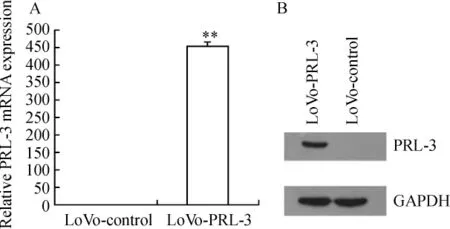
Figure 1.The expression of PRL-3 in LoVo-PRL-3 cells and LoVo-control cells.±s.n=3.**P<0.01 vs control.A:PRL - 3 mRNA was determined by realtime PCR.The results show that expression level of PRL-3 mRNA in LoVo-PRL-3 cells was 430-fold higher than that in LoVo-control cells.B:Cell lysates(30 μg)from stable cell lines were used to confirm PRL-3 expression by immunoblotting with anti-PRL-3 antibody.The expression level of PRL-3 protein in LoVo-PRL-3 cells was significantly increased but not detected in LoVo-control cells.图1 LoVo-PRL-3和LoVo-control细胞PRL-3表达水平比较
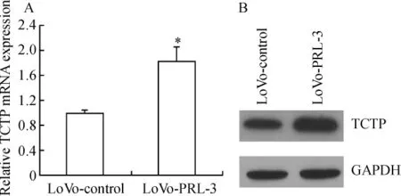
Figure 2.The expression of TCTP in LoVo-PRL-3 cells and LoVo-control cells.±s.n=3.*P<0.05 vs control.A:TCTP mRNA was determined by real- time PCR.The results show that expression level of TCTP mRNA in LoVo-PRL-3 cells was 1.83-fold higher than that in LoVo - control cells.B:Western blotting analysis of TCTP protein level.Cell lysates(30 μg)from stable cell lines were used to confirm TCTP expression by immunoblotting with anti-TCTP antibody.The expression level of TCTP protein in LoVo-PRL-3 cells was significantly increased compared with LoVo-control cells.图2 LoVo-PRL-3和LoVo control细胞TCTP表达水平比较
2 TCTP-siRNA有效抑制LoVo-PRL-3细胞TCTP mRNA和蛋白的表达
Real-time PCR结果显示,转染TCTP-siRNA 24 h、48 h和72 h后能显著抑制TCTP mRNA在Lo-Vo-PRL-3细胞中的表达(P<0.01),见图3A。Western blotting结果显示,在转染TCTP-siRNA 48h和72h后 LoVo-PRL-3细胞 TCTP蛋白的表达受到显著抑制(P<0.01),见图3B。
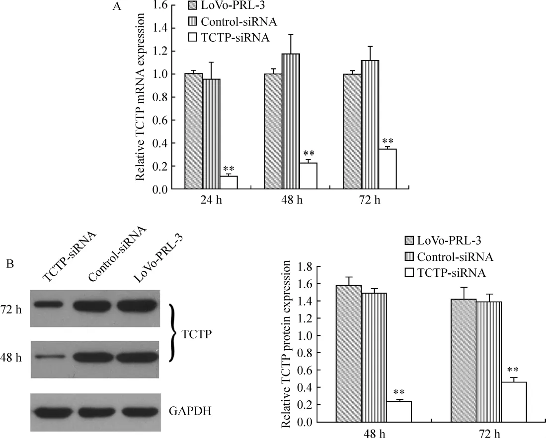
Figure 3.Gene silencing of TCTP by siRNA transfection.±s.n=3.**P<0.01 vs LoVo-PRL-3 or control-siRNA.A:The level of TCTP expression in LoVo-PRL-3,control-siRNA and TCTP-siRNA cells were examined 24,48 and 72 h after siRNA transfection using quantitative real-time PCR.B:Western blotting also showed that 48 and 72 h after transfection with siRNA,LoVo-PRL-3 cells transfected with TCTP-siRNA dramatically down-regulated the expression of TCTP protein.图3 siRNA干扰抑制TCTP表达
3 siRNA干扰TCTP表达抑制PRL-3引起的细胞增殖
如图4所示,与对照细胞LoVo-control相比,转染PRL-3后细胞的增殖能力显著增强(P<0.05),siRNA干扰TCTP表达后又能显著抑制PRL-3引起的细胞增殖(P<0.05)。
4 siRNA干扰TCTP表达抑制PRL-3引起的细胞迁移和侵袭
如图5所示,与对照细胞LoVo-control相比,转染PRL-3后细胞的迁移和侵袭能力显著增强(P<0.05),siRNA干扰TCTP表达后又能显著抑制PRL-3引起的细胞迁移和侵袭(P<0.01)。
讨 论
结直肠上皮组织的恶性转变及其进一步的转移是由多种分子共同参与完成的[10]。因此发现并研究肿瘤转移过程中的重要相关分子及其信号通路对于预防和治疗肿瘤转移具有重要意义。已有大量研究表明PRL-3在肿瘤细胞转移过程中起到重要作用[11]。目前认为,PRL-3促进肿瘤转移主要通过促进肿瘤细胞增殖、促进肿瘤细胞迁移和侵袭及促进血管形成等方面起作用。已有研究发现PRL-3高表达与卵巢癌的进展相关,并在体外通过RNA干扰方法抑制卵巢癌细胞的PRL-3表达能显著抑制细胞增殖[4]。Zeng等[11]发现 PRL -3 转染至无转移潜能的中国仓鼠卵巢细胞(Chinese hamster ovary cell,CHO cell)后,CHO细胞的迁移和侵袭能力明显增强,并在裸鼠模型中形成转移灶。此外,通过RNA干扰方法抑制具有转移能力的结肠癌细胞DLD-1的内源性PRL-3表达后能明显抑制其在小鼠体内的转移成瘤能力[12]。同时,已有研究表明,PRL -3表达增高的转移瘤细胞可以在血管内萌芽生长[13]。在侵润性乳腺癌的脉管系统中发现PRL-3的表达上调[14]。综上所述,PRL-3促进肿瘤转移是多种分子参与的多方面作用的结果。因此,发现并研究PRL-3促肿瘤转移过程中的关键性分子对于预防和治疗肿瘤转移具有重要意义。
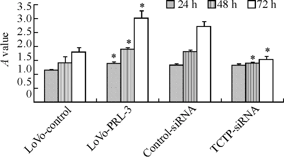
Figure 4.Knockdown of TCTP inhibited the PRL-3-promoted proliferation of LoVo cells.±s.n=3.*P<0.05 vs LoVo-control or control-siRNA.The LoVo-control and LoVo-PRL-3 cells(5×10³cells/well)were cultured in 96-well plates.After 24h,LoVo-PRL-3 cells were transfected with 100 nmol/L TCTP-siRNA or control-siRNA.Then,24,48 and 72h after transfection,cell proliferation was measured.Results show that elevated PRL-3 expression in LoVo cells promoted cell growth compared with LoVo-control cells,while knockdown of up-regulated TCTP expression in LoVo-PRL-3 cells led to suppression of cell growth.图4 siRNA干扰TCTP表达抑制PRL-3引起的细胞增殖
TCTP是一类广泛存在于动植物细胞中在序列上高度保守且同源性很高的蛋白家族。在TCTP与肿瘤相关功能研究中,Tuynder等[15]发现,TCTP在逆转的白血病、乳腺癌、结肠癌、肺癌和黑色素瘤细胞中均显著下调,通过反义cDNA或siRNA干扰抑制肿瘤细胞TCTP表达,会导致肿瘤细胞恶性表型的抑制。更重要的是,最近有研究发现,TCTP在前列腺癌和结肠癌细胞转移的不同阶段:细胞增殖、细胞凋亡、细胞迁移和侵袭及转移均具有重要作用[8,9]。此外,已有研究表明,编码TCTP蛋白的基因TPT1受转录因子CREB的调控[16]。转录因子CREB的活化需要其第133个丝氨酸残基的磷酸化,细胞外信号调节激酶1/2(ERK1/2)就是引起它第133个丝氨酸残基磷酸化的激酶之一[17]。而PRL-3可以使整合素β1磷酸化水平下调,从而引起ERK1/2磷酸化水平的增加从而发挥促进肿瘤转移作用[18]。因此,我们猜想PRL-3可能通过上调TCTP来促进肿瘤细胞转移。
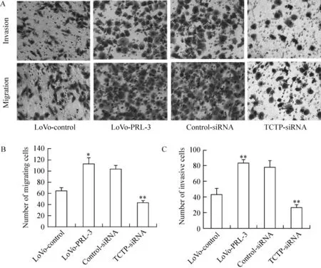
Figure 5.Down-regulation of TCTP by RNAi attenuated the PRL-3-promoted migration and invasion activity of LoVo cells.±s.n=3.**P <0.01 vs LoVo-control or control-siRNA.A:LoVo-PRL-3 cells were treated with TCTP-siRNA specific for TCTP or control-siRNA.Forty-eight hours after transfection,an aliquot of 1×10⁵cells was placed in upper chamber with 0.1 mL serum-free medium,whereas the lower chamber was loaded with 0.6 mL of medium containing 10%fetal bovine serum.After 20 h of incubation,the cells migrating or invading to the underside of filter were stained and observed under a microscope(×200).B,C:Statistical plots of the number of cells invading the Transwell membranes by migration assay and matrigel invasion assay.Results showed that PRL - 3 promoted the migration and invasion of LoVo cells,while knockdown of up-regulated TCTP expression in LoVo-PRL-3 cells led to suppression of migration and invasion.图5 siRNA干扰TCTP表达抑制PRL-3引起的细胞迁移和侵袭
为了证实TCTP蛋白在PRL-3促进肿瘤转移过程中的重要作用,我们首先将PRL-3基因转染到内源性低表达PRL-3的结肠癌细胞株LoVo中,并检测LoVo-PRL-3细胞及未转染PRL-3的对照细胞LoVo-control的TCTP表达。结果发现TCTP mRNA和蛋白在转染PRL-3后分别上调1.8倍和2.1倍。之后,为了证实TCTP的上调在PRL-3促肿瘤转移中的作用,我们通过siRNA干扰抑制LoVo-PRL-3细胞中上调的TCTP表达。结果发现,抑制TCTP表达能显著降低PRL-3引起的细胞增殖、迁移和侵袭能力。因此我们认为,TCTP是PRL-3促结肠癌转移过程中的重要分子,PRL-3通过上调TCTP表达促进结肠癌细胞增殖、迁移和侵袭;用siRNA靶向抑制TCTP表达可能是预防和治疗结肠癌转移的有效手段。
[1]Kim KA,Song JS,Jee J,et al.Structure of human PRL -3,the phosphatase associated with cancer metastasis[J].FEBS Lett,2004,565(1 -3):181 -187.
[2]Saha S,Bardelli A,Buckhaults P,et al.A phosphatase associated with metastasis of colorectal cancer[J].Science,2001,294(5545):1343 -1346.
[3]Miskad UA,Semba S,Kato H,et al.Expression of PRL -3 phosphatase in human gastric carcinomas:close correlation with invasion and metastasis[J].Pathobiology,2004,71(4):176-184.
[4]Polato F,Codegoni A,Fruscio R,et al.PRL -3 phosphatase is implicated in ovarian cancer growth[J].Clin Can cer Res,2005,11(19 Pt 1):6835 -6839.
[5]Schwering I,Brauninger A,Distler V,et al.Profiling of Hodgkin's lymphoma cell line L1236 and germinal center B cells:identification of Hodgkin's lymphoma-specific genes[J].Mol Med,2003,9(3 -4):85 -95.
[6]Wang L,Peng L,Dong B,et al.Overexpression of phosphatase of regenerating liver-3 in breast cancer:association with a poor clinical outcome[J].Ann Oncol,2006,17(10):1517-1522.
[7]Bommer UA,Thiele BJ.The translationally controlled tumour protein(TCTP)[J].Int J Biochem Cell Biol,2004,36(3):379 -385.
[8]Gnanasekar M,Thirugnanam S,Zheng G,et al.Gene silencing of translationally controlled tumor protein(TCTP)by siRNA inhibits cell growth and induces apoptosis of human prostate cancer cells[J].Int J Oncol,2009,34(5):1241-1246.
[9]Ma Q,Geng Y,Xu W,et al.The role of translationally controlled tumor protein in tumor growth and metastasis of colon adenocarcinoma cells[J].J Proteome Res,2010,9(1):40-49.
[10]崔 翼,汪建平,彭俊生,等.结肠癌的蛋白质组学研究[J].中国病理生理杂志,2008,24(5):1013-1017.
[11]Zeng Q,Dong JM,Guo K,et al.PRL -3 and PRL -1 promote cell migration,invasion,and metastasis[J].Cancer Res,2003,63(11):2716 -2722.
[12]Kato H,Semba S,Miskad UA,et al.High expression of PRL-3 promotes can cer cell motility and liver metastasis in human colorectal cancer:a predictive molecular marker of metachronous liver and lung metastases[J].Clin Cancer Res,2004,10(21):7318 -7328.
[13]Guo K,Li J,Tang JP,et al.Catalytic domain of PRL - 3 plays an essential role in tumor metastasis:formation of PRL -3 tumors inside the blood vessels[J].Cancer Biol Ther,2004,3(10):945 -951.
[14]Parker BS,Argani P,Cook BP,et al.Alterations in vascular gene expression in invasive breast carcinoma[J].Cancer Res,2004,64(21):7857 -7866.
[15]Tuynder M,Fiucci G,Prieur S,et al.Translationally controlled tumor protein is a target of tumor reversion[J].Proc Natl Acad Sci U S A,2004,101(43):15364 -15369.
[16]Andree H,Thiele H,Fahling M,et al.Expression of the human TPT1 gene coding for translationally controlled tumor protein(TCTP)is regulated by CREB transcription factors[J].Gene,2006,380(2):95 -103.
[17]Shaywitz AJ,Greenberg ME.CREB:a stimulus-induced transcription factor activated by a diverse array of extracellular signals[J].Annu Rev Biochem,1999,68:821 -861.
[18]Peng L,Xing X,Li W,et al.PRL -3 promotes the motility,invasion,and metastasis of LoVo colon cancer cells through PRL-3-integrin beta1-ERK1/2 and-MMP2 signaling[J].Mol Cancer,2009,8:110.